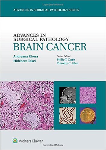
 (Part of the series on Advances in Surgical Pathology)
Editors: Andreana Rivera, MD and Hidehiro Takei, MD.
Series Editors: Philip T. Cagle, MD and Timothy C. Allen, MD
Publisher: Wolters Kluwer | Lippincott, Williams & Wilkins – 257 pages
Book Review by: Nano Khilnani
Brain cancer is a very rare disease affectingly only one to two percent of all those who contract cancer in the United States. But the level of morbidity (severity of sickness) is significant among those who have it. And the mortality rate is also quite high, meaning that only a few among those who get this disease, survive.
According to the Central Brain Tumor Registry of the United States (CBTRUS), between 2007 and 2001, there were 115,799 people (7.25 among 100,000) who had malignant tumors in the brain and central nervous system (CNS). There were also 227,376 people (14.17 among 100,000) who had benign cases. So the total for malignant and benign cases was 343,175. For this five-year period, the average per year comes to 68,635 for both malignant and benign cases.
While the annual average (in the five year period 2007-2011) for malignant brain tumor cases is only 23,160 in a population of around 320 million people, every person who is diagnosed with it has to go through a period of pain and suffering, and a high number die from it. The editors of this book write that for the patients, as well as for all family members, “primary brain cancers exert a devastating impact” on them.
Brain cancer is not easily identifiable. There are “130 histologic brain tumor entities” based on the World Health Organization classification system for this disease. The editors point out: “the diagnosis is often not straightforward because brain tumors are highly heterogeneous neoplasms that can originate from cells within the brain parenchyma, mininges, or nerves.”
Identifying and tracking biologic behavior is also not easy in brain tumors. It ranges from benign to highly malignant. The editors point out “even benign tumors can be lethal due to their location in the brain, their ability to infiltrate locally, and their propensity to undergo malignant transformation.”
Adding to this difficulty is the fact that “each tumor entity is associated with its own biology, treatment, and prognosis. Combined, these unique features make brain cancer pathologic diagnosis and treatment very challenging.”
For these and other reasons, this book on brain cancer is essential to own and study closely, for anyone who is already involved in, or plans to get into this medical specialty. It has been put together by outstanding teachers and practitioners in the area of brain tumor pathology.
Except for one person who is a research scientist, the other 24 contributors to this book are all doctors of medicine who are practicing pathologists, radiologists or surgeons, or are professors of these medical specialties at various institutions. Among the 25 people who provided material for this book, 18 are based in the United States, five in Japan, and two in South Korea.
The material in this book has been allocated into 19 chapters organized around five sections:
- Overview:
- Normal Histology
- Histopathology
- Diffuse (Infiltrating) Gliomas
- Circumscribed Astrocyctomas
- Ependymomas
- Other Neuroepithelial Tumors
- Neuronal / Mixed Glioneuronal Tumors
- Embryonal Tumors
- Choroid Plexus Tumors
- Pineal Region Tumors
- Sellar and Suprasellar Region Tumors
- Meningeal Tumors
- Hereditary Syndromes
- Tumors of Cranial and Peripheral Nerves and Other Tumors
- Hematolymphoid and Histiocytic Tumors
- Central Nervous System Metastasis
- Imaging
- Correlation of Imaging with Pathology
- Molecular Pathology
- Glial Tumors
- Nonglial Tumors
- Current Therapy and Prognosis
- Brain Cancer Treatment
Here are some of the benefits you will derive from this book:
- Effectively interpret the most challenging histopathology findings with in-depth coverage of basic diagnostic features, differential diagnoses, and diagnostic pitfalls.
- Recognize key diagnostic characteristics with the aid of hundreds of high-quality color illustrations.
- Make optimal use of immunohistochemistry and molecular testing as they apply to diagnosis, prognosis, and treatment.
- Gain a holistic view of the field with a clinicopathologic overview of brain tumors, the correlation between imaging and pathology, and look at the current state of therapeutics.
Editors:
Andreana Rivera, MD is Staff Neuropathologist in the Department of Pathology and Genomic Medicine at Houston Methodist Hospital in Houston, Texas.
Hidehiro Takei, MD is Associate Professor in the Department of Pathology and Laboratory Medicine at Weill Medical College of Cornell University in New York, New York; and Pathologist in the Department of Pathology and Genomic Medicine at Houston Methodist Hospital in Houston, Texas.
Series Editors:
Philip T. Cagle, MD is Professor of Pathology and Laboratory Medicine at Weill Medical College of Cornell University in New York, New York; and Director of Pulmonary Pathology at Houston Methodist Hospital in Houston, Texas.
Timothy Craig Allen, MD is Professor in the Department of Pathology, and Director of Anatomic Pathology at the University of Texas Medical Branch in Galveston, Texas.


