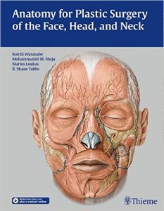
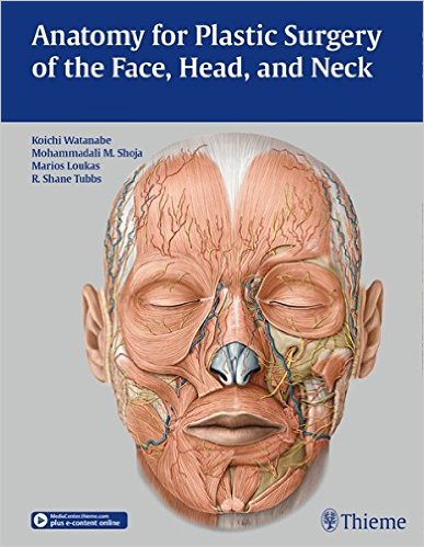
 Editors: Koichi Watanabe, MD, Mohammedali M. Shoja, MD, Marios Loukas, MD, and R. Shane Tubbs, PhD
Publisher: Thieme – 226 pages
Book Review by: Nano Khilnani
This book fills a gap. Most anatomy books do not provide sufficiently detailed information (discussions and graphics) on regional anatomy for plastic surgeons working on the face, head and neck. On the other hand, in most facial, head and neck plastic surgery books, anatomy is not emphasized and anatomical details provided are insufficient for the surgeon to expertly complete the procedures.
The editors write that the reason for this shortcoming is that the anatomy in the head and neck region, especially that applicable to plastic surgery, is still developing, and anatomical textbooks often do not provide the most up-to-date information.
In this book, “we have attempted to include the latest anatomical understanding of the head and neck anatomy from a plastic surgeon’s perspective,” the four editors named above write in their Preface. They inform us that the anatomy of the head and neck is “extremely complicated” and it is not the same inside the head of every person (italics mine). And, they write further: “the details differ among individuals and during different stages of life.”
Among the anatomical differences in particular individuals are the following:
- Thickness of the tissues
- Their changes in response to aging
- Anatomical variations in vessels, nerves, and muscles
Most anatomical atlases do provide detailed images of any given anatomical region. Learning, retaining the knowledge, and being able to visualize it is essential for success in surgery. The views in atlases however, are two-dimensional
The editors emphasize the importance for surgeons and surgeons-to-be, of having thorough knowledge of, and being able to visualize the three-dimensional anatomy of the area of the human body they are specializing in, and operating on, not just two-dimensional anatomy. “Novice surgeons typically memorize the two-dimensional image of their surgical field. Because of this, surgical results are sometimes unsatisfactory, or surgical complications may occur,” they write.
“To perform surgeries with a high degree of difficulty, a surgeon has to be able to vividly visualize the three-dimensional regional anatomy of the surgical field,” they emphasize.
Thirty-nine specialists in various fields, including aesthetic surgery, anatomy, basic sciences, developmental biology, cosmetic surgery, dentistry, gerodontology, gerontology, head and neck surgery, maxillofacial surgery, neuroradiology, neurological surgery, oculoplastic and orbital surgery, ophthalmology, oral biology, otorhinolaryngology, pediatric neurosurgery, plastic and reconstructive surgery, from nine countries – Canada, Germany, Japan, Qatar, South Korea, Turkey, the United Kingdom, the United States, and West Indies – authored the 21 chapters of this book, which are:
- Neurocranium and Facial Skeleton
- Anterior Skull Base
- Middle-Skull Base
- Soft Tissue of the Scalp and Temporal Regions
- Arterial Supply of the Facial Skin
- Arteries of the Face and Neck
- Veins of the Face and Neck
- Facial Nerve and Temporal Bone
- Peripheral Branches of the Facial Nerve
- Sensory Nerves of the Head and Neck
- Superficial Musculoaponeurotic System and the Facial Soft Tissues
- Mimetic Muscles
- Orbital Anatomy
- Orbital Soft Tissues
- Eyelid Anatomy
- Nasal Cavity and Paranasal Sinuses
- External Nose
- Auricle and External Acoustic Meatus
- Mandible and Masticatory Muscles
- Oral Cavity and Pharynx
- Neck
Videos
In addition to the material in the book, you can also view five informative videos on www.MedicalCenter.Thieme.com. When prompted during the process of registering your copy of this book, simple enter the access code found on the inside from cover of your book. These videos are:
- Facial muscles and facial nerve on the anterior face: lower face, middle face
- Dissection of the external nose: muscles on the external nose, bony and cartilaginous structure
- Main trunk of the facial nerve and branches: landmarks of the facial nerve trunk, temporal branch, zygomatic branch, buccal branch, marginal mandibular branch, cervical branch
- Sensory nerves of the face: supraorbital nerve, infraorbital nerve, zygomaticofacial nerve, mental nerve
- Layers of the temporal region: superficial temporal fascia, deep temporal fascia, temporalis muscle
This book provides highly detailed views of the complex and highly varied anatomy of the face, head and neck for plastic and otorhinolaryngology surgeons and surgical residents. It empowers and thus enables them to perform procedures with a high degree of success, and avoid mistakes and mishaps. This book overcomes the shortcomings of others in this field.
Editors:
Koichi Watanabe, MD, PhD is Assistant Professor in the Department of Anatomy at Kurume University School of Medicine in Fukuoka-Prefecture, Japan
Mohammedali M. Shoja, MD is Research Scientist in the Section of Pediatric Neurosurgery at Children’s Hospital in Birmingham, Alabama.
Marios Loukas, MD, PhD is Dean of Basic Sciences, Professor and Chair in the Department of Anatomical Sciences at St. Georges University in Grenada, West Indies.
Shane Tubbs, MS, PA-C, PhD is Professor and Chief Scientific Officer at Seattle Science Foundation in Seattle, Washington.


