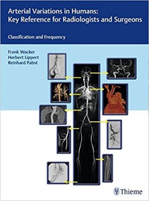
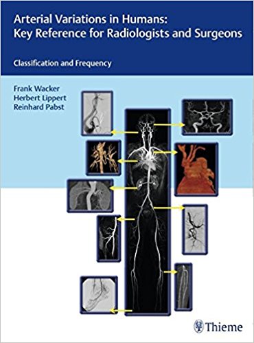
 Editors: Frank Wacker, MD; Herbert Lippert MD; and Reinhard Pabst, MD
Publisher: Thieme – 209 pages, with 924 illustrations
Book Review by: Nano Khilnani
About 95 percent of arteries in our bodies follow what is known to be normal anatomical structure and branching. But for some arteries the normal pattern is shown in as few as only 30 percent of patients.
Rarely mentioned in standard anatomy textbooks are these variations – the deviations in a given artery’s origin, its topographical localization, and the area it supplies blood to. This book fills that gap of knowledge.
This is a large (9”x12”) volume with more than 900 images, averaging almost four hand a half per page, so there is a lot to learn about the details of the vascular anatomy of the human body, as shown and discussed in its 47 chapters.
What is starkly unusual about this work is that each chapter presents actual images of arterial and venous structures in patients that are different than their normal patterns, accompanied by full-color sketches. Some of the differences are of the following types: abnormal structure or branching with larger or smaller than normal diameters of some vessels, additional vessels connecting from one part to another, anomalous structures, and malformed vessels.
Fourteen specialists (all from Germany except one from Ireland) on the anatomy and physiology of blood vessels and nerves, diagnostic and interventional radiology, and general radiology authored the chapters of this volume. They are organized around five Parts that we name below to give you a bird’s eye view of what you will find in this atlas that includes discussions of the functions of the vessels and their interrelationships.
- Part I. Heart and Thorax
- Part II. Pelvis and Abdomen
- Part III. Lower Limbs
- Part IV. Upper Limbs
- Part V. Head and Spinal Cord
This early (2017) release of a 2018 book is an enhancement of the 1985 work published by Drs. Lipper and Pabst entitled Arterial Variations in Man – Classification and Frequency. With the collaboration of Dr. Frank Wicker and his team, the original atlas has been expanded and includes in this edition, angiograms obtained with computed tomography (CT), digital subtraction angiography (DSA), and magnetic resonance imaging (MRI).
While CT scanning was invented in 1972 by British engineer Godfrey Hounsfeld, DSA by three people named Castellanos, Robb, and Steinberg in the 1970s, and MRI in 1970 by medical doctor and research scientist Raymond Damadan, their development as technologies enabling the viewing of blood vessels in bony, dense soft tissue and other environments did not develop until much later. As is the case with many inventions, initial versions do not have sophisticated and detailed capabilities, that typically emerge later.
The three editors point out that what they learned about the structure and variations in arterial anatomy in their rotations as residents in angiography and interventional radiology were much more diverse than what they had learned earlier in their textbooks and classes in medical school.
This book depicts variations in arterial structure and delivery of blood supply to organs that can potentially save lives. How? Through at least two ways, the editors point out:
- Correct detection and interpretation of pathologic changes requires knowledge of both the normal and anomalous arterial blood supply.
- Under certain circumstances some variations in blood supply can have negative effects. Such knowledge is critical in therapy planning including surgery after endoscopic examination and interventional radiology. Unpleasant surprises can be avoided with correct, detailed visualization prior to these actions.
Detailed knowledge of vasculature (both of arteries and veins) is extremely important. Below we mention two ways wherein lack of such knowledge could have severe negative effects:
- A superficial brachial artery does not a direct effect on the function of the forearm and hand. But if that artery is mistaken for a vein and should thiobarbiturate be injected into it, severe necrosis of the hand would result.
- When selective trans-arterial chemo-mobilization or radio-embolization is used for the treatment of hepatic cancer, even small aberrant hepatic arteries can cause significant side-effects due to off-target embolization.
In addition to variations in anatomical structure and branching, some arteries are malformed. Malformations can have negative effects as well, as for example if both coronary arteries originate from a pulmonary artery.
This is a very important book that any physician should own, whether they have, or have not encountered examples of variant vasculature in their practices. It is a much-awaited expanded and enhanced edition of a truly pioneering effort that begun in 1985.
Editors:
Frank Wacker, MD is Professor and Director of the Diagnostic and Interventional Radiology at Hannover Medical School in Hannover, Germany.
Herbert Lippert, MD is Professor at the Institute of Anatomy at Hannover Medical School in Hannover, Germany.
Reinhard Pabst, MD is Professor at the Institute of Anatomy at Hannover Medical School in Hannover, Germany.


