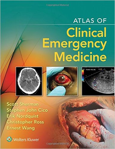
 Editors: Scott C. Sherman, MD; Stephen John Cico, MD; Erik Nordquist, MD; Christopher Ross, MD; and Ernest Wang, MD
Publisher: Wolters Kluwer | Lippinocott, Wilkins & Williams – 374 pages
Book Review by: Nano Khilnani
This is a highly visual book, using one, two, three, and sometimes more images on each page. We all know that “a picture is worth a thousand words,” as has been expressed numerous times. But many of us probably do not know that in seven out of 10 times, we process information mainly in visual format (pictures and words) as opposed to other means such as hearing and touching. This has been revealed and documented in numerous research studies.
Dr. Scott C. Sherman makes this point in the Preface, and additionally, he writes that he used a camera as an important tool in educating himself daily during his emergency medicine residency, beginning in 2001, at a community hospital on the northwest side of Chicago.
For the most part, the images used in this book are full-color photographs of the injury, abnormality, condition, discomfort, disease, or disorder that brought the patient to the emergency department of the hospital.
But besides color photos, many other means and types of imaging are used, including but not limited to: charts, elastography, electrocardiography (ECG), electroencephalography (EEG), magnetic resonance imaging (MRI), magneto-encephalography (MEG), nuclear medicine, radiographs, sketches, thermography, tomography (including computed tomography or CT imaging) ultrasound, and x-rays.
Some 275 doctors from all over the United States authored the 340 chapters of this book. More precisely, these are not chapters but rather, topics. Each topic is presented on a single page with images and text. The book is organized around 15 sections, namely:
- Section A: Trauma (22 chapters)
- Section B: Neurologic (17 chapters)
- Section C: Ophthalmologic (28 chapters)
- Section D: Ear, Nose, Throat (19 chapters)
- Section E: Dental (8 chapters)
- Section F: Cardiovascular (22 chapters)
- Section G: Respiratory (8 chapters)
- Section H: Gastrointestinal (17 chapters)
- Section I: Genitourinary (14 chapters)
- Section J: Obstetrics/Gynecology (6 chapters)
- Section K: Dermatologic (45 chapters)
- Section L: Orthopedic (75 chapters)
- Section M: Pediatrics (38 chapters)
- Section N: Toxicologic (7 chapters)\
- Section O: Environmental (14 chapters)
The presentation of material is quite consistent, straightforward and simple, and such simplicity makes for easier and quicker learning. In each section, the topics are presented alphabetically. Such type of presentation makes this book easy to use as a reference guide, and saves much time, a precious commodity in critical medical emergency situations.
Below the images on each page, you will find the same three subheadings with bullet points:
- Clinical Presentation
- Diagnosis
- Management
Consistency is even revealed in the use of same colors in the text box for each of the above subheadings: blue, brown and green, respectively.
Let us see the type of content you will find in this book by taking a look at one of the topics covered. In Section A – Trauma – let us take a look on page 7 on topic 5, Emergency Department Thoracotomy.
The photo at the top of the page, with the caption Repair of a cardiac injury, shows a surgeon working on a patient’s heart.
In the blue Clinical Presentation textbox, you are informed that Emergency Department (ED) thoracotomy (an incision into the area of the chest that has the heart, thoracic aorta, lungs, anterior spine, esophagus, etc.) is “a drastic final effort to save the life of an arresting trauma victim. The primary goals of the procedure are:
- Hemorrhage control
- Release of cardiac tamponade (blood, clots, fluid, gas, and/or pus)
- Facilitation of internal / open cardiac massage
- Prevention of air embolism (gas bubbles)
- Exposure of the descending thoracic aorta for cross-clamping, and
- Repair of cardiac and pulmonary injury
This is followed by the brown Diagnosis textbox in which you learn that for thoracotomy there are two accepted indications:
- Penetrating chest trauma with signs of life in the field: traumatic arrest with previously witnessed cardiac activity, and unresponsive low hypotension (systolic blood pressure or BP below 70 mm Hg)
- Blunt chest trauma with signs of life lost in the emergency department: besides systolic BP below 70, rapid exsanguination (blood loss) from the chest tube: over 1,500 mL
Lastly, in the green Management textbox, you are instructed to:
- Assess throughout the thoracotomy procedure, the cause of the cardiac arrest and the interventions performed to correct the cause of that arrest
- Start the procedure on the left side for quicker access to the pericardium. Incision is performed from the sternum to the posterior axillary line in the fourth or fifth intercoastal space. After cutting through deep tissues, ribs are spread and lung is pushed to access the pericardium
Pericardiotomy is performed followed by inspection of myocardium for lacerations. Cardiac defects can be covered with a finger until the wound can be closed with sutures, staples, or insertion of a Foley catheter.
- Cardiac massage is performed with one or both hands to provide improved cardiac output. Defibrillation (15 to 30 J) is performed with internal paddles placed on either side of the heart.
The entire contents of the print edition are available for download as an eBook. Follow these simple steps:
- Go to http://solution.lww.com/access
- Enter the Access Code found on the inside front cover of this book
- Enter your information, click Submit, and follow the on-screen instructions to start reading your eBook
Your book purchase includes not only a complimentary download of the enhanced eBook for iOS, Android, PC and Mac, but also these features:
- Complete content with enhanced navigation
- Powerful search tools and smart navigation cross-links that pull results from content in the book, your notes, and even the web
- Cross-linked pages, references, and more for easy navigation
- Highlighting tool for easier reference of key content throughout the text
- Ability to take and share notes with friends and colleagues
- Quick reference tabbing to save your favorite content for future use
This is an excellent volume on clinical emergency medicine. It is authoritative because so many (275) doctors competent in various specialties related to emergency medicine contributed content for it. It is easily accessible because of its excellent organization and presentation of material. And it is highly utile because it has just the essential information needed to make the right decision in an emergency setting.
Editors:
Scott C. Sherman, MD is Associate Residency Director at Cook County Emergency Medicine Residency, and Associate Professor of Emergency Medicine at Rush Medical College in Chicago, Illinois.
Stephen John Cico, MD, MEd is Associate Professor of Clinical Emergency Medicine and Pediatrics and Director of the Pediatric Emergency Medicine Fellowship Program in the Division of Emergency Pediatrics at Indiana University School of Medicine. He is also Attending Physician in Pediatric Emergency Medicine in the Department of Emergency Medicine at Emergency Medicine and Trauma Center at Riley Hospital for Children in Indianapolis, Indiana.
Erik Nordquist, MD is Assistant Program Director of Cook County Emergency Medicine Residency, and Professor of Emergency Medicine at Rush Medical College in Chicago, Illinois.
Christopher Ross, MD, FRCPC, FACEP, FAAEM is Associate Professor of Emergency Medicine and Associate Chair of Professional Education in the Department of Emergency at John H. Stroger, Jr. Hospital of Cook County in Chicago, Illinois.
Ernest Wang, MD, FACEP is the Alvin H. Baum Family Fund Chair of Simulation and Innovation at North Shore University Health System in Evanston, Illinois. He is also Assistant Dean for Medical Education, and Clinical Professor in Emergency Medicine at University of Chicago Pritzker School of Medicine in Chicago, Illinois.


