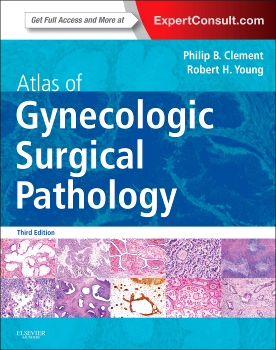
 Editors: Philip B. Clement, MD, and Robert H. Young, MD, FRCPath
Publisher: Elsevier Saunders – 568 pages
Book Review by: Nano Khilnani
There are certain terms that are used frequently in this textbook, and although medical students and practitioners may not only be familiar with them, but may also have seen various, and even numerous versions of them, we provide brief definitions and descriptions below, obtained from several online medical lexicons. We do this so the reader can get a clearer understanding of the contents found in this book
Some of these terms are: carcinoma, hyperplasia, lesion, neoplasm, pathology, and tumor
A carcinoma (a common term for cancer) is any of various types of malignant neoplasm derived from epithelial cells, chiefly glandular (adenocarcinoma) or squamous (squamous cell carcinoma); the most commonly occurring kind of cancer.
A hyperplasia is an increase in the number of normal cells in a tissue or organ, excluding tumor formation, whereby the bulk of the part or organ may be increased.
A lesion is an injured or diseased spot or area on or in the body
A neoplasm is an abnormal tissue that grows by cellular proliferation more rapidly than normal and continues to grow after the stimuli that initiated the new growth cease. Neoplasms show partial or complete lack of structural organization and functional coordination with the normal tissue, and usually form a distinct mass of tissue that may be either benign (benign tumor) or malignant (cancer).
Pathology is the medical specialty practice concerned with all aspects of disease, but with special reference to the essential nature, causes, and development of abnormal conditions, as well as the structural and functional changes that result from the disease processes.
A tumor is any swelling or inflammation on or in the body; an inflamed tissue.
This book, the first edition of which was published in 2000, and the second edition in 2008, continues in this third edition published in 2014 to be an easy-to-use a guide to the study, diagnosis, and treatment of lesions (which includes cancer, neoplasm and tumor) of the female reproductive system.
It contains numerous actual photos of abnormal and diseased tissue, as well as microscopic images of them in its 20 chapters. The amount of information found in it is voluminous. This book is one of the most authoritative sources of information and insight in its field, written by two experienced pathologists.
Here is an overview of the subjects covered in this valuable book:
- Non-neoplastic lesions and benign tumors of the vulva
- Malignant tumors of the vulva
- The vagina
- Tumor-like lesions and benign tumors of the uterine cervix
- Invasive squamous cell carcinoma of the cervix and its precursors
- Adenocarcinomas of the cervix, related tumors, and their precursors
- Non-neoplastic lesions of the uterine corpus
- Endometrial hyperplasia and carcinoma
- Mesenchymal and mixed epithelial-mesenchymal tumors of the uterine corpus and cervix
- Trophoblastic lesions, miscellaneous primary uterine neoplasms, hematopoietic neoplasms, and metastic neoplasms to the uterus
- The fallopian tube and broad ligament
- Tumor-like lesions of the ovary
- Surface epithelial-stromal tumors, general features, serous tumors, and mucinous tumors
- Surface epithelial-stromal tumors, endometrioid, clear-cell, transitional, squamous, rare, undifferentiated, and mixed cell types
- Germ cell tumors of the ovary
- Sex cord-stromal and steroid cell tumors of the ovary
- Miscellaneous primary ovarian tumors
- Metastic tumors of the ovary (including pseudomyxoma peritonei, hematolymphoid neoplasms, and tumors with functioning stroma)
- Endometriosis and lesions of the second mullerian system
- Tumor-like lesions and tumors of the peritoneum (excluding mullerian lesions)
To access the complete contents of this textbook online – which are fully searchable – and to use the other valuable features, simply visit the website indicated below, enter the Activation Code by scratching off the sticker on the inside front cover of this book, and follow the instructions online to activate your access.
If you are already registered, login at www.expertconsult.com, scratch off the Activation Code on the inside front cover of this book, enter it into the Add a Title box, click on Activate Now, and click the title under My Titles.
If you’re a first-time user, click on Register Now, fill in your user information and click Continue, then activate your book by entering your Activation Code into the Enter Activation Code box; then click on Activate Now, and click the title under My Titles.
An additional resource is available to purchasers of this book on www.ClinicalKey.com
Go to the inside back cover of this text, and you will find that ClinicalKey is designed to serve you well by providing you these important core components:
- Comprehensive Content – The most current, evidence-based answers available for every medical and surgical specialty.
- Trusted Answers – Content supplied by Elsevier, the world’s leading provider of health and science information.
Unrivaled Speed to Answer – Faster, more relevant clinical answers, so you can spend less time searching and more time caring for patients.
This is an outstanding and comprehensive textbook on its subject, authored by its two authors named below.
Philip B. Clement, MD is Consultant Pathologist in the Department of Pathology at Vancouver General Hospital, and Emeritus Professor of Pathology at the University of British Columbia in Vancouver, Canada.
Robert H, Young, MD, FRCPath is Pathologist in the James Homer Wright Pathology Laboratories at Massachusetts General Hospital, and Robert E. Scully Professor of Pathology at Harvard Medical School in Boston, Massachusetts.


