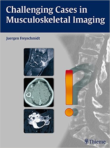
 Author: Juergen Freyschmidt, MD
Publisher: Thieme – 371 pages, with 1,438 illustrations
Book Review by: Nano Khilnani
This could be called a ‘look-and-learn’ book, with more than 1,400 images to look at, and learn by closely reading and understanding the text on the pages, as well as accompanying captions. The total number of images is fairly large for a book of this length. If you divide that number by the pages, it works out to almost four images per page.
Nearly all of the images are machine scans of one type or another, including: computed tomography (CT), Magnetic Resonance Imaging (MRI), positron emission tomography (PET), radiography, or ultrasound. An unusual type of scanning is also used here, rarely found in other medical books with machine scans: it is called bone scintigraphy including single-photon emission computed tomography (SPECT).
Dr. Freyshmidt has delved quite deeply into the subject of imaging bone and muscle in all major parts of the human body, in this unusual and very important book. This has been written for students and practitioners in orthopedics, pathology, radiology, and other areas, and there is much to learn here. This large-size volume is definitely a keepsake if you are in, or plan to be in these and related medical specialties. It is certainly worth the investment to own it. Here is a brief overview of what you will find in it:
- From Symptom to Diagnosis
- Selecting the Correct Image Modality
- Image Interpretation
- Skull
- Sclerotic Changes
- Osteolytic Lesions and Lesions with Mixed Features
- Spine
- Mono- and Bisegmental Changes
- Oligo- and Multisegmental Changes
- Diseases of Spinal Entheses and Joints
- Sacrum
- Pelvis
- Sclerotic Changes
- Osteolytic Changes and Changes Associated with Decreased Bone Density
- Unusual Fractures
- Bone Lesions with a Predominantly Extraosseous Component
- Soft-Tissue Mineralization
- Hip Region
- Shoulder Girdle and Thoracic Cage
- Clavicle
- Scapula
- Ribs
- Sternum
- Anterior Chest Wall as a Whole
- Upper Limb
- Upper Arm
- Forearm
- Hands (Bone and Joint Disease)
- Lower Limb
- Equivocal MRI Findings
- Predominantly Osteolytic Changes
- Predominantly Osteoclerotic Changes
- Changes with Mixed Features
- Extraosseous Lesions
The author cautions that knowledge of, and experience in radiology, isn’t enough in correctly interpreting images to discover abnormalities, disorders, and diseases in patients. What he strongly recommends that you do, in order to arrive at a “clinically useful diagnosis” is to use a systematic approach to analyzing images. By piecing together data derived from the images, you can arrive at correct conclusions, he suggests.
He writes: “By contrast, ‘old hands’ at radiology can diagnose most cases at a glance owing to their extensive experience but this ‘shooting from the hip’ will often miss the mark when it comes to more challenging diagnoses. Thus, having made several blunders in the past, I have made it a practice to recheck a seemingly obvious diagnosis for its logical plausibility and correctness.”
With this book, Dr. Freyschmidt aims to show how challenging cases can be diagnosed simply by using a systematic approach to interpreting images. He emphasizes that many cases that were initially difficult to interpret were later found to be not difficult after using a systematic approach. He discusses the subject of image interpretation in section 1.2 of the first chapter.
Each chapter has the following headings followed by discussions:
- Case No. and Fig. No. (referencing images)
- Case Description (in a boxed area)
- Radiologic Findings
- Location
- Pathoanatomic Background of the Findings
- Assignment to a Possible Basic Entity
- Synopsis and Discussion
- Final Diagnosis and Comments
This is a well-laid out book that is easy to read and understand, and unique because the author takes a systematic approach at analyzing and interpreting images, and arriving at a diagnosis of the disease or disorder.
Author:
Juergen Freyschmidt, MD is Professor in the Reference Center for Osteoradiology at Zentralklinikum Bremen Witte, University Teaching Hospital of the University of Gottingen in Bremen, Germany.

