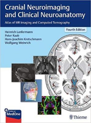
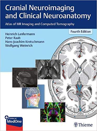
 Editors: Heinrich Lanfermann, Peter Raab, Hans-Joachim Kretschmann, Wolfgang Weinrich
Publisher: Thieme – 513 pages
Book Review by: Nano Khilnani
This large-format (about 13” x 9”) neurology book contains a tremendous wealth of information within its five Parts that contains 11 chapters with subject-related knowledge, plus two additional chapters entitled ‘Specimens and Technique’ and ‘Bibliography’. All this is organized within 513 pages containing a lot of illustrations, numbering 930.
But just to not scare you with what you’re probably thinking is a huge amount of data to study, and then, to understand their effects on the human brain, I’m saying don’t be scared because this is really just an atlas with much of the space occupied by images of various types.
So it is actually something to keep as a reference book or manual to look up when you want to find answers to specific questions or solutions to specific problems.
We list the chapter titles below so that you can have a broad idea of what you will find in this large-sized reference source:
- Part I – Introduction
- Introduction
- Tomography and Landmarks
- Part II – Atlas
- Coronal Sections
- Saggital Sections
- Transverse Sections
- Brainstem
- Part III – Topography of the Head and Neck
- Topography of the Cranium, Intracranial Spaces, and Contained Structures
- Facial Topography
- Topography of the Head-Neck Region
- Nervous System – Neurofunctional Systems and Neuroactive Substances
- Neurofunctional Systems
- Neurotransmitters and Neuromodulators
- Part V – Appendix
- Specimens and Technique
- Bibliography
Additional information is available to you when you register your copy of this textbook online. Go to: www.MedOne.Thieme.com/9783136726044. Then, input the access code found by scratching off the film found on the inside front cover of this book, or scan the QR code found there.
This fourth (2019) English edition of the book contains many updates of information in the previous edition, as well as new information. This edition contains:
- Adaptations of axial sections to conventional CT angulation sparing the orbits
- Descriptions of the development of cross-sectional imaging have been trimmed
- Detailed illustrations created by Professors Krectschmann and Weinrich are based on the analogous transfer of anatomical cranial sections to high-resolution graphics
- Elaborations on cranial vessels and neuro-functional systems
- New chapters on brain maturation and the petrous part of the temporal bone
The main aims of this book are the following:
- Depiction of brain anatomy in the atlas section of this book in the three conventional planes used in cross-sectional imaging.
- Graphic rendition of principal arterial territories in the supra- and infratentorial regions.
- Description and imaging of the principal neuro-functional systems in multi-planar slices.
- Elucidation of the relationship of the topography of neuro-functional systems with the site of a lesion and its clinical symptoms
This book very well performs these objectives and in our opinion, it is a valuable, highly authoritative resource for all who intend to specialize in neurology and neurosurgery, as well as for those who are well-established in this field.
Editors:
Heinrich Lanfermann, MD is Professor and Director of the Department of Diagnostic and Interventional Neuroradiology at Hanover Medical School in Hanover, Germany.
Peter Raab, MD is a member of the Senior Staff and Head of Neuroradiological Oncologic Imaging at the Institute of Diagnostic and Interventional Neuroradiology at Hanover Medical School in Hanover, Germany.
Hans-Joachim Kretschmann, MD is Professor and former Director of the Department of Neuroanatomy at Hanover Medical School in Hanover, Germany.
Wolfgang Weinrich, MD is Professor and former Head of the Neurological Clinic at Nordstadt Hospital in Hanover, Germany.
Contributors:
Eva Bultmann, MD is a member of the Senior Staff, and Head of Neuroradiological Pediatric Imaging in the Department of Diagnostic and Interventional Neuroradiology at Hanover Medical School in Hanover, Germany.
Anja Glesermann, MD is affiliated with Department of Diagnostic and Interventional Neuroradiology at Hanover Medical School in Hanover, Germany.
Dina Witfoth, MSc, PhD is affiliated with Department of Diagnostic and Interventional Neuroradiology at Hanover Medical School in Hanover, Germany.


