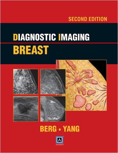
Editors: Wendie A. Berg, MD; and Wei Tse Yang, MD
Publisher: Amirsys – Wolters Kluwer | Lippincott, Williams & Wilkins
Book Review by: Nano Khilnani – 800 pages, plus other sections
In 2012, 1.68 million cases of breast cancer cases were reported worldwide, according to an article in Wikipedia. Of these, 31 percent or 522,000 resulted in deaths, about twenty percent of which were found to have cancer through screening. Fortunately, survival rates in England and the United States are high, with between 80 and 90 percent living for five years after detection and treatment.
This is likely due to the widespread availability of healthcare personnel and facilities in these countries. Survival rates are also probably high in other parts of the developed world, but in less developed areas, outcomes for those who contract breast cancer, are likely quite dim.
For women worldwide, cancer of the breast is the leading cancer, with about one in four of them having this type of cancer, and the other three having cancer in other parts of their bodies.
What are some of the common risk factors for breast cancer? Among them are: female sex, drinking alcohol, early age at first menstruation, family history, having children late in life, hormone replacement therapy during menopause, ionizing radiation, lack of physical exercise, and older age.
While this book is not focused on breast cancer but on imaging modalities used in diagnosing the breast for abnormalities, anomalies, diseases, and disorders of this organ and surrounding areas, a good portion of it deals with breast imaging which can reveal lesions and tumors that are either benign or malignant, and other discoveries.
For those who have a malignancy, the treatments options are typically chemotherapy, hormonal therapy, or radiation therapy, and in extreme cases, surgery, including mastectomy or removal of the breast(s).
Twenty-seven physicians and other specialists involved in teaching, research, and treatment related to the breast wrote the chapters of this book, which we present below as an overview for you, of what you can study in this book. The authors are mainly imaging specialists, oncologists, professors, radiologists, and surgeons.
- Section 1. Anatomy and Normal Variants:
- Anatomy
- Normal Variants
- Section 2. Imaging
- Modalities
- Section 3. Interpretive Criteria
- Introduction and Overview
- Interpretive Criteria
- Section 4. Lesion Imaging Characteristics
- Masses
- Calcifications, Typically Benign
- Calcifications, Not Typically Benign
- Additional Findings
- Ultrasound Lesion Characteristics
- MR Findings
- Section 5. Histopathologic Diagnoses
- Introduction and Overview
- Benign Lesions
- Risk Lesions
- Locally Aggressive Lesions
- Malignant Lesions
- Section 6. Signs and Symptoms
- Introduction and Overview
- Nipple
- Skin
- Axilla
- Breast
- Vascular Entities
- Section 7. Postoperative and Post-Treatment Imaging
- Introduction and Overview
- Results of Benign Intervention
- Results Following Treatment for Malignancies
- Section 8. Special Topics
- Hormonal Changes
- Pregnancy
- Infections and Inflammation
- Breast Manifestations and Systemic Conditions
- Trauma
- Male Breast
- Artifacts
- Section 9. Screening Malignancies
- Issues in Screening
- Breast Cancer Treatment
- Treatment Planning
- Section 10. Procedures
- Image-Guided Procedures
- Surgical Procedures
You will be pleased to learn that you can access the contents of this print book online! To access your Amirsys eBook Advantage:
- Scratch off the silver coating on the inside front cover of this book to reveal the license key
- Go to http://amirsys.com online
- Register your title with this license key
This website is for individual use only. For additional details, please read the License Agreement available on the registration page. For technical assistance, please contact [email protected]
Amirsys eBook Advantage Features:
- Searchable content
- Hundreds of additional images
- Continuous updates
- Expanded diagnostic tips and references
This book, like others in the Amirsys/Lippincott series on diagnostic imaging, provides information in outline and bullet-point format, which enables you the reader to make a general diagnosis in less time, a precious element in the field of medicine.
Another plus in using this book is the very large number and variety of images available in black-and-white and in color.
Each topic in each Section contains a Key Facts box with four parts, namely:
- Terminology
- Imaging
- Pathology
- Clinical Issues
These are followed by detailed presentations of each of these parts. A Differential Diagnosis (with Malignancies, Risk Lesions, and Benign Lesions) follows Imaging, and a list of Selected References follows Clinical Issues. Detailed captions of the images provide important details crucial to correct diagnosis and interpretation. In some instances, a Diagnostic Checklist is provided following Clinical Issues.
One of the new features in this second edition is the Introduction and Overview in some Sections. For example in Section 6, Signs and Symptoms, discussions on the important topics are provided:
- Palpable Mass
- Pain, Erythema
- Axillary Adenopathy
- Axillary Nodal Calcifications
- Nipple Discharge, Nipple Retraction, Nipple Inversion, Paget Disease
- Skin Retraction, Invasion, or Thickening
- Shrunken Breast
This is an outstanding book on diagnosing breast diseases. The textual and visual materials are presented in a fashion that enables readers to quick grasp the features of abnormalities, and look into details as well to better understand them. The types of imaging used are many, among them: computed tomography (CT), magnetic resonance imaging (MRI), tomosynthesis, ultrasound, and x-rays.
Editors:
Wendie A. Berg, MD is Professor of Radiology at University of Pittsburgh School of Medicine and works at Magee-Women’s Hospital of UPMC in Pittsburgh, Pennsylvania.
Wei Tse Yang, MD is Professor of Radiology, Chief of the Section of Breast Imaging, and Medical Director of the Ben and Julie Rogers Breast Diagnostic Clinic at the University of Texas MD Anderson Cancer Center in Houston, Texas.


