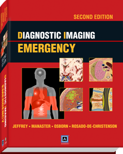
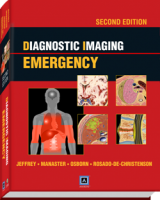
 Editors: R. Brooke Jeffrey, MD, B.J. Manaster MD, PhD, FACR, Anne G. Osborn, MD, FACR, and Melissa L. Rosado-de-Christenson, MD, FACR
Publisher: Amirsys | Lippincott, Williams & Wilkins – 1,010 pages
Book Review by: Nano Khilnani
The contents of this book are available in searchable form at: http://ebooks.amirsys.com.
You can access the Amirsys ebook Advantage features found on that website by scratching off the silver coated tab found on the inside front cover to reveal the license key. If you’re an existing user you can log in to your account; if you’re a new user you can register for a new account. Both types of users can register this book with their license key.
Once on the site noted above, you can search for the content you are looking for. You can also get to an expanded section of diagnostic tips and references, and additional images.
The four editors of this volume received material from 55 authors – most of them MDs, a few PhDs, and some holders of the MBBS degree.
The contents in this book of over a thousand pages are simply organized into Part I (Trauma) and Part II (Nontrauma). Each of these two Parts has four Sections, namely: Central Nervous System, the Chest/Cardiovascular System, the Abdominal Pelvic System, and the Musculoskeletal System.
Each Section of the book in Part I and Part II of this volume contains numerous images of patients’ human parts that are damaged, injured, or affected by a disease or disorder. Sections begin with an Introduction and discussions of different topics.
For example, Section 2, Chest/Cardiovascular in Part I (Trauma), starts with an Introduction to Chest Imaging, Trauma. The Section then has discussions on:
• Airway/Lung: Tracheobronchial Laceration, Pulmonary Contusion/Laceration
• Mediastinum/Cardiovascular: Pneumomediastinum, Hemopericardium, Pneumo-pericardium, Traumatic Aortic Injury, Esophageal Rupture
• Pleura/Chest Wall/ Diaphragm: Pneumothorax, Hemothorax, Rib Fracture, Flail Chest, Sternal Fracture, Spinal Fracture, Diaphragmatic Rupture
• Pediatric: Child Abuse
Some of the Imaging Modalities discussed in this Section 2 are: Chest Radiography, Chest CT, Ultrasound, Angiography, and Magnetic Resonance Imaging or MRI.
Discussions follow on the parts of the chest or thoracic area, which include: the chest wall, the pleura, lungs, airways, the mediastinum, the diaphragm, and the possible use of life support devices.
Each of the topics in the section is covered in a way that is different than found in other medical books. A text box on the topic of Tracheobronchial Laceration presents Key Facts, with the subtopics: Terminology, Imaging, Top Differential Diagnoses, and Clinical Issues.
It defines tracheobronchial laceration as the “disruption of trachea or bronchi following blunt or penetrating injury.” The type of Imaging to be used in this type of injury would be either Radiography or CT. The Top Differential Diagnoses would be of Pneumomediastinum, Pneumothorax, Esophageal Rupture and Esophageal intubation.
The clinical issues to be addressed in this type of injury would be:
• Signs and symptoms: respiratory distress, continuous air leak despite chest tube draining, and extensive subcutaneous gas
• 30% of affected patients die; 50% of fatalities occur within one hour after trauma
• Delayed diagnosis is common: 70% not identified in 1st 24 hours; 1 month delay in 40%
• Airway stenosis, atelectasis, mediastinitis sepsis
• Diagnosis is confirmed with bronchoscopy
• Treatment: prompt surgical repair‘
‘
The information provided in the text box is followed by presentation of various images showing injuries. For example, a chest radiograph taken after a stab at the neck shows a wound sustained by the victim.
A tracheal laceration was found on him during surgery. Another image shows a large right pneumothorax with the atelectic right lung “falling away” from the mediastinum secondary to a right mainstem bronchus laceration. The “falling lung” sign denotes tracheobronchial injury.
The presentation of images as some examples of tracheobronchial laceration is followed by a detailed outline of the topics presented in the Key Facts text box. This detailed outline now includes considerations relating to Pathology, and a Diagnostic Checklist. A Selected References list is also provided for further study.
This is a very well organized and laid out book with facts that can be easily digested and images that actually show the reader the injuries of patients so he or she can see what a tracheobronchial laceration, for example, looks like, even though it is shown in an x-ray image.
The editors named below have done a fantastic job in gathering and presenting the vast amount of material contained in this valuable volume. Congratulations are in order to them, and to the contributors. And we must not forget those who did other jobs such as text editing, image editing, medical editing, illustrations, art direction and design, and the individuals who were the Lead Editor and the Publishing Lead.
R. Brooke Jeffrey, MD is Professor and Vice Chairman in the Department of Radiology at Stanford University School of Medicine in Stanford, California.
B.J. Manaster, MD, PhD, FACR is Professor of Radiology at the University of Utah School of Medicine in Salt Lake City, Utah.
Anne G. Osborn, MD, FACR is University Distinguished Professor, Professor of Radiology, and holds the William H. and Patricia W. Child Presidential Endowed Chair in Radiology at the University of Utah School of Medicine in Salt Lake City, Utah.
Melissa L. Rosado-de-Christenson, MD, FACR is Section Chief, Thoracic Imaging, in Saint Luke’s Hospital in Kansas City, and Professor of Radiology at University of Missouri-Kansas City in Kansas City, Missouri.

