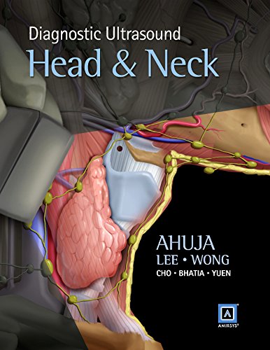
 Author: Anil T. Ahuja, MBBS;
Contributing Authors: Yolanda Y.P. Lee, MBChB; K.T. Wong, MBChB; Carmen C.M. Cho, MBBS; Kunwar S.S Bhatia, B Med Sci; H.Y. Yuen, MBChB; Stella Sin Yee Ho, RDMS
Publisher: Elsevier–Amirsys – 576 pages, 1000+ images
Book Review by: Nano Khilnani
Ultrasound, also known as ultrasonography, is one of several means of imaging used in looking at structures within the human body, diagnosing diseases and disorders, and deciding on the ideal means of treatment.
Other ways of medical imaging are: computed tomography, known in short as CT; elastography, electroradiography or ECG; electroencephalography or EEG; endoscopy, and magnetic resonance imaging or MRI.
Lesser-used methods are: magnetoencephalography or MEG; medical photography, nuclear medicine functional imaging techniques such as positron emission tomography; photoacoustic imaging, tactile imaging, and thermography.
This book is useful for practitioners and students of endocrinology, oncology, radiology, sonography, surgery, and all those involved in diagnosing and treating diseases in the head and neck.
It focuses on ultrasound and is a combination of three previously-published books:
- Diagnostic Imaging: Ultrasound
- Diagnostic and Surgical Imaging Anatomy: Ultrasound
- Expert: Ultrasound
It is divided into three parts and eleven sections:
Anatomy
- Head and Neck
Diagnoses
- Introduction and Overview
- Thyroid and Parathyroid
- Lymph Nodes
- Salivary Glands
- Lumps and Bumps
- Vascular
- Post-Treatment Change
- Intervention
Differential Diagnoses
- Head and Neck
- Thyroid and Parathyroid
Each section has subsections that further look into other parts of the human anatomy. Materials presented in the subsections are organized around these segments:
- Terminology
- Imaging Anatomy
- Anatomy Imaging Issues
- Related references
You can access the entire contents of this book plus additional materials through Amirsys eBook Advantage. Do the following:
- Scratch off silver coating on the inside front cover of your book to get your license key
- Go to: http://ebooks.amirsys.com
- Existing users: log in to your account
- New eBook Advantage users: Register for a new account
- Register your title with this license key
(You may need to log out and log back in to see your new eBook in your list of registered titles)
This is a wide-ranging compilation of more than a thousand ultrasound images of the human anatomy, with a variety of numerous patterns of disease and disorders in the head and neck. Along with full-color photos of patients with abnormal conditions (e.g. abscesses, cutaneous openings, floppy eyelids, lumps, masses, and swelling) are presented black-and-white images that “look inside” those patients.
The following authors are to be congratulated for this outstanding book:
Author:
Anil T. Ahuja, MBBS (Bom), MD (Bom), FRCR, FHKCR, FHKAM (Radiology) is Professor of Imaging and Interventional Radiology, and Chairman of the Department of Imaging and Interventional Radiology at Prince of Wales Hospital. He is also on the Faculty of Medicine at the Chinese University of Hong Kong in Hong Kong (SAR), China.
Contributing Authors:
Yolanda Y.P. Lee, MBChB, FRCR, FHKCR, FHKAM (Radiology) is Associate Consultant and Clinical Associate Professor (Honorary) in the Department of Imaging and Interventional Radiology at the Prince of Wales Hospital, and is on the Faculty of Medicine at the Chinese University of Hong Kong in Hong Kong (SAR), China
K.T. Wong, MBChB, FRCR, FHKCR, FHKAM (Radiology) is Consultant and Clinical Associate Professor (Honorary) in the Department of Imaging and Interventional Radiology at the Prince of Wales Hospital, and is on the Faculty of Medicine at the Chinese University of Hong Kong in Hong Kong (SAR), China
Carmen C.M. Cho, MBBS, FRCR, FHKCR, FHKAM (Radiology) is Associate Consultant and Clinical Assistant Professor (Honorary) in the Department of Imaging and Interventional Radiology at the Prince of Wales Hospital, and is on the Faculty of Medicine at the Chinese University of Hong Kong in Hong Kong (SAR), China
Kunwar S.S. Bhatia, B Med Sci, BMBS, MRCS, DLO, FRCR is Assistant Professor (Honorary) in the Department of Imaging and Interventional Radiology at the Prince of Wales Hospital, and is on the Faculty of Medicine at the Chinese University of Hong Kong in Hong Kong (SAR), China
H.Y. Yuen, MBChB, FRCR, FHKCR, FHKAM (Radiology), MPH is Associate Consultant and Clinical Associate Professor (Honorary) in the Department of Imaging and Interventional Radiology at the Prince of Wales Hospital, and is on the Faculty of Medicine at the Chinese University of Hong Kong in Hong Kong (SAR), China.
Stella Sin Yee Ho, RDMS, RVT, PhD is Adjunct Associate Professor in the Department of Imaging and Interventional Radiology at the Prince of Wales Hospital, and is on the Faculty of Medicine at the Chinese University of Hong Kong in Hong Kong (SAR), China.
Simon S.M. Ho, MBBS, FRCR is Clinical Associate Professor (Honorary) in the Department of Imaging and Interventional Radiology at the Prince of Wales Hospital, and is on the Faculty of Medicine at the Chinese University of Hong Kong in Hong Kong (SAR), China.


