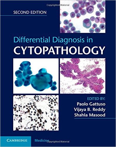
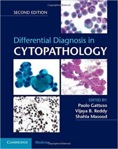
 Editors: Paolo Gattuso, MD; Vijaya B. Reddy, MD; and Shahla Masood, MD
Publisher: Cambridge University Press – 681 pages, with 1,000+ macrographs
Book Review by: Nano Khilnani
In recent years, minimally invasive diagnosis and treatment has been gradually replacing excisions (biopsies) and open surgery. This is just one in a number of many new ways to deliver personalized medical services more efficiently and effectively, resulting in better outcomes in healthcare in the United States. The editors – Drs. Gattuso, Reddy, and Masood – also point out that “integrated care has resulted in better treatment planning and improved patient satisfaction.”
But a lot of improvements in healthcare delivery in the U.S. would not have been possible without ongoing medical research and innovation that has the enabled the development of better technological devices and tools to diagnose, treat, and provide prognosis for a large number of abnormalities, anomalies, conditions, diseases and disorders in people.
The editors point out importantly in their Preface that pathologists – those who study disease processes, and cytopathologists – who trace the disease processes in cells – have played a central role in improving healthcare in this country, with their work in the development, validation, and implementation of those advanced technologies. Cytopathologists have been actively engaged in learning more about the morphologic features of diseases that develop in the cell and their diagnosis, including diagnostic pitfalls.
These days, cytologic preparations are used routinely in ancillary studies to get the most accurate information about the specific biology and nature of a particular disease process. Cytopathology is an exciting field for the potential it offers of a discovery that could save millions of lives. But it is also challenging because research is a long-drawn process and an important life-changing discovery does not happen without putting in a lot of effort behind it.
The editors write that a good cytopathologist must also be a good surgical pathologist, they write: “in many ways a good cytopathology practice is similar to looking through the keyhole and being able to see the entire room.”
This book has been developed for use by practicing pathologists, including anatomic pathologists and other specialists with pathology, cytopathologists, cytotechnologists. It is an ideal resource for residents in pathology and fellows in cytopathology, as well as the academic community.
Thirty-three contributors including the three editors named above – who are professors and practitioners of several specialties within cytology and pathology, as well as genomic science, laboratory medicine, and radiology, from mainly the United States, but also from Canada and Saudi Arabia – contributed content primarily by authoring its 19 chapters named below:
- The PAP smear
- Exfoliative pulmonary cytology
- Body Cavity fluids
- Gastrointestinal tract cytology
- Urine cytology
- Cerebrospinal fluid and intraocular cytology
- Cytopathology of the central nervous system
- Fine-needle aspiration of the thyroid gland
- Parathyroid gland, and head and neck cytopathology
- Cytopatholgy of the salivary glands
- Cytopathology of the breast
- Fine-needle aspiration of lymph glands
- Fine-needle aspiration of lungs, pleura, and mediastinum
- Fine-needle aspiration of the liver
- Fine-needle aspiration of the pancreas
- Fine-needle aspiration of soft tissue and bone
- Fine-needle aspiration of the kidney and adrenal gland
- Cytology of the gonads
- Fine-needle aspiration cytology of tumors of unknown origin
You as purchaser of this book can access online the entire contents of this book: text and images, which are fully searchable. Here’s how you set up your Cambridge Books Online account:
- Go to http://ebooks.Cambridge.org/register and Click Register
- Enter your details and click Submit
- You will receive a confirmation email. Please follow the instructions in that email to activate your registration
To activate your access code using your Cambridge Books Online account:
- Scratch off the Activation Code found on the inside from cover of your book
- Go to http://ebooks.Cambridge.org and click Login, followed by User Login
- Log in using your Cambridge Books Online account
- Click on My Bookshelf
- Enter your Activation Code and click on Activate Now
- Click on the book’s title to view the book’s content
The materials in each chapters are presented in a systematic manner that typically contain the following elements:
- Outline – a box with bulleted points, at the beginning of each chapter
- Topics and subtopics with bullet points throughout each chapter
- Under the topics: clinical and cytologic features, special stains, diagnostic techniques
- Differential diagnosis in light blue boxes with bullet points, below topics and subtopics
- Pearls in smaller boxes in light blue boxes with bullet points, below topics and subtopics
- Micrographs in full color with detailed captions
- References
This is a book with detailed coverage of cytopathologic diseases within several organ systems, using a focused bullet-point system. It uses one of the currently most widely-used approaches to diagnosing diseases at the basic cellular unit level, which is cytologic evaluation of exfoliative and fine-needle aspiration.
This is not only an excellent but essential text for practitioners and trainees in general pathology, as well as specialists in cytopathology and cytotechnology at all levels of their careers.
Among the key features of this encompassing book in cytopathology are:
- It address a comprehensive variety of benign and neoplastic conditions
- It utilizes a consistent structure with bullet-point text for quick access and assimilation
- It presents a full spectrum of infectious and inflammatory disorders in detail
- Its information is not limited to light microscopic findings but includes many other genetic, molecular, immunologic diagnostic modalities
- It gives you the clinical and diagnostic criteria needed when formulating a diagnosis and differential diagnosis
Editors:
Paolo Gattuso, MD is the Otho S.A. Professor and Director of Anatomic Pathology at Rush Medical College in Chicago, Illinois.
Vijaya B. Reddy, MD is Professor and Associate Chair of the Department of Pathology at Rush Medical College in Chicago, Illinois.
Shahla Masood, MD is Professor and Chair of the Department of Pathology and Laboratory Medicine at the University of Florida College of Medicine in Jacksonville, Florida.

