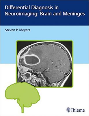
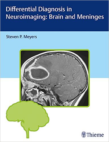
 (Part of a three-volume series. The other two are: Differential Diagnosis in Neuroimaging: Head and Neck, and Differential Diagnosis in Neuroimaging: Spine.
Author: Steven P. Meyers, MD
Publisher: Thieme – 646 pages, with 1952 illustrations
Book Review by: Nano Khilnani
This is a definitive guide on identifying and examining diseases in the brain, authored by the renowned neuro-radiologist Dr. Steven Meyers, who has had about 30 years of hands-on experience researching, studying and teaching about neurological disorders to medical students, residents and fellows in neurology, neurosurgery, orthopedics, otolaryngology, radiology and related medical specialties.
This large and thick book catalogues and examines various brain pathologies based on neuro-imaging results and locations. The numerous scans obtained through several imaging technologies are presented in this volume. As most of you know. the most widely-used imaging modalities are: computed tomography (CT), positron emission tomography (PET) and a combination of these two methods (PET/CT).
The large quantity of contents of this book are presented essentially in tabular form laid out in chapters sandwiched between an Introduction and References. There are 26 chapters in this book that are organized around five Sections we list below. Next to each Section we write the chapter/table titles to give you an overview of what is covered in this book
- Brain (Intra-Axial Lesions) – congenital and histogenic malformations of the brain; supratentorial solitary intra-axial mass lesions; solitary intra-axial lesions in the posterior cranial fossa (infratentorial); multiple intra-axial lesions in the brain; multiple or diffuse lesions involving white matter in children; bilateral lesions involving the basal ganglia and/or thalami; neuro-degenerative disorders; ischemia and infarction involving the brain and / or brainstem in adults; ischemia and infarction involving the brain and / or brainstem in children; intrasellar and juxtasellar lesions; lesions in the pineal region
- Ventricles and Cisterns – lateral ventricles-common masses; common third ventricle masses; fourth ventricle lesions; excessively small ventricles; dilated ventricles; abnormal or altered configurations of the ventricles; solitary intraventricular lesions in children; solitary intraventricular lesions in adults; contrast-enhancing ventricular margins
- Extra-Axial Lesions – solitary extra-axial mass lesions; multifocal extra-axial lesions
- Meninges – abnormalities involving the dura; multifocal and/or diffuse leptomeningeal abnormalities
- Vascular Abnormalities – congenital and developmental vascular anomalies/variants; acquired vascular disease
The goal of this book and the other two books in this series is “to present the imaging features of neuroradiological abnormalities in an easy-to-use resource, with extensive utilization of figures for illustration,” in the words of the author.
All the tables in which the neurological abnormalities, anomalies, conditions, diseases and disorders are laid out, have three columns:
- Lesions
- Imaging Findings
- Comments
Detailed captions are provided next to the presented images to impart information on the findings, in order to determine the next correct course of action.
For example in chapter 3 entitled Intra-axial lesions in the posterior cranial fossa (infratentorial, in Fig 1.116 on page 97, the two images are of an 18-month-old male with a diffuse astrocytoma in the pons. Graphic (a) shows an intermediate signal on sagittal T1-weighted imaging, and Graphic (b) has a high signal on axial FLAIR. The tumor enlarges the pons and compresses the fourth ventricle. What needs to be done? The text provides the available options, with the pros and cons of each choice.
This is a highly valuable book that contains a tremendous amount of information in words and pictures. It should be a must-have guide and reference manual for anyone involved in neurology and neurosurgery.
Author:
Steven P. Meyers, MD , PhD, FACR is Professor of Radiology / Imaging Sciences, Neurosurgery and Otolaryngology at the University of Rochester Medical Center, and Director of the Radiology Residency Program at the University of Rochester School of Medicine and Dentistry in Rochester, New York.


