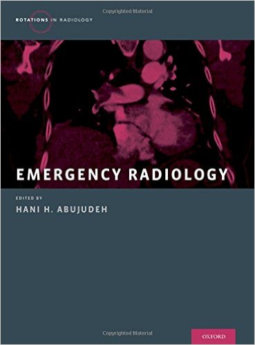
Editor: Hani H. Abujudeh, MD
Publisher: Oxford University Press – 415 pages
Book Review by: Nano Khilnani
Emergency Radiology is a new subspecialty of radiology that enables its practitioners to image parts of the anatomy that are injured and that need to be examined promptly in order to decide on the immediate course of action to be taken by the physician-surgeon in the emergency room of the hospital.
It is a combined study of organs, organ systems and a range of various imaging modalities. Its essential nature embodies multi-organ, multi-modality views. The knowledge and skills acquired in this field enable the practitioner to present to acute-care providers, an immediate, comprehensive evaluation of the patient’s injury or injuries to begin surgery or perform other forms of treatment to mitigate the victim’s physical condition.
From a real-life occupational perspective, emergency radiologists provide time-critical support to trauma surgeons and other personnel in emergency rooms. In some cases, they also provide tele-radiology and off-hour services to institutions worldwide that lack such qualified personnel.
Fifty-four specialists in imaging services, radiology, surgery, and other fields, from the United States (mainly Massachusetts), India, Saudi Arabia, Thailand, and Venezuela wrote the 66 chapters of this book that are organized around nine Sections, namely:
- Section I. Head, Neck, and Spine Emergencies
- Section II. Chest Emergencies
- Section III. Abdominal Emergencies
- Section IV. Pelvic Emergencies
- Section V. Bone: Lower Extremity Emergencies
- Section VI. Bone: Upper Extremity Emergencies
- Section VII. Pediatric Emergencies
- Secrion VIII. Special Topics in Emergency Radiology
- Section IX. Nuclear Medicine Emergencies
Among the imaging techniques emergency radiologists must master are: computed tomography (CT), magnetic resonance imaging (MRI), position emission tomography (PET) and x-ray. There has been growing demand for qualified and competent emergency radiologists in recent years who are able to correctly interpret images.
One of the challenges emerging in the field of emergency radiology is growing demand for them in the effort to provide better healthcare services with more accurate interpretation of the imaging results. The others are:
- Greater demand for increasing manpower, with an aging population in the United States
- Development of evidence-based best practices to provide care in patient-centered effective and efficient ways
- Defining the scope of the specialty for board certification, credentialing, and other quality assurance practices
The materials in all the chapters are systematically organized, and in standard ways. You the reader can learn about each subject more easily and quickly when your expectation is predictable
For example in chapter 1, Traumatic Brain Injury, as in other chapters, you will see the following headings after the chapter title and bylines in the order given below:
Definition
Clinical Features
Anatomy and Physiology
How to Approach the Image (different types of images and findings presented in this section)
What Not To Miss
Differential Diagnoses
Imaging Findings
Clinical Issues
Key Points
Further Reading
Let’s take a brief look at some of the facts and discussions presented in the first two headings of this chapter:
Definition: One of the most common causes of neurological disability in the United States is traumatic brain injury (TBI) which affects 1.7 million people in this country each year. It results in approximately 275,000 hospitalizations, around 52,000 deaths, and it has a very high cost of care: about $76 billion.
Clinical Features: This major type of injury to the brain results in cerebral contusion, diffuse axonal injury (DAI), intracranial hemorrhage (ICH), and in severe cases, coma and death. The degree of the injury is classified as mild, moderate, or severe based on these four factors: duration of the loss of consciousness, Glassgow Coma Scale (GCS) score, presence of post-traumatic amnesia lasting less than an hour, and no definite findings on standard CT or MR scans.
This is an excellent book that is authoritative with more than fifty specialists’ contributions, thorough with more than 66 chapters on a large range of topics, and with materials (text and images) that are well organized, presented and written.
Editor:
Hani J. Abujudeh, MD, MBA, FSIR, FACR is Associate Professor of Radiology at Massachusetts General Hospital and Harvard Medical School in Boston, Massachusetts. He is also Chairman of Radiology at Cooper University Hospital and Cooper Medical School at Rowan University in Camden, New Jersey.


