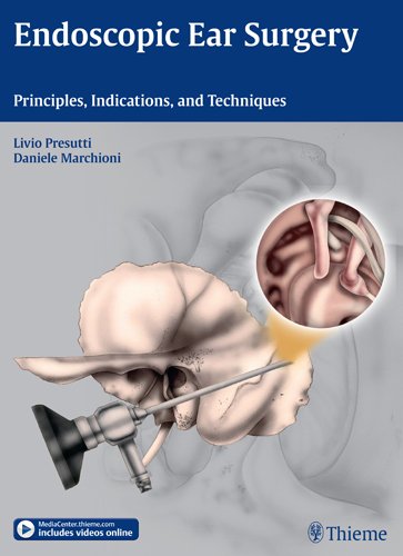
 Editors: Livio Presutti, MD; and Daniele Marchioni, MD
Publisher: Thieme – 403 pages with 951 illustrations
Book Review by: Nano Khilnani
This outstanding book on surgery of the middle and inner ear, with large (9” x 12”) pages, contains numerous graphics, including full drawings and photos of various types of tissue, diagnostic machines, organs, and surgical instruments; micrographs, sketches, and x-rays.
Each page has anywhere from a single photograph to composites of as many as a dozen photos. All this makes for easier absorption of material for you the student or practitioner of otology, the specialty on the ear, particularly the middle and inner ear space.
An article on middle ear endoscopy in the e-medicine section of www.medscape.com briefly gives us the anatomy of the human ear as follows:
The human ear is composed of external, middle or tympanic section having the malleus, incus, and stapes; and inner or labyrinth section with the semicircular canals, vestibule, and cochlea portions.
The auricle and external acoustic meatus or external auditory canal, compose the external ear. The external ear functions to collect and amplify sound, which then gets transmitted to the middle ear.
The tympanic cavity (middle ear) extends from the tympanic membrane to the oval window and contains the bony conduction elements of the malleus, incus, and stapes. The primary functionality of the middle ear is that of bony conduction of sound via transference of sound waves in the air collected by the auricle to the fluid of the inner ear.
The inner ear, also called the labyrinthine cavity, is essentially formed of the membranous labyrinth encased in the bony osseus labyrinth. The labyrinthine cavity functions to conduct sound to the central nervous system (our brain) as well as to assist in balance.
The study of the anatomy, function, examination of disease, and surgery in this part of the head is done with the aid of the endoscope. The editors of this volume – Dr. Livio Presutti and Dr. Daniele Marchioni – write that for a period of about 30 years (period not specified but probably from 1967 to 1996) no progress was made in EES, short for endoscopic ear surgery.
Mer was the first to describe the use of an endoscope to view the detailed anatomy, with small dimensions, of the middle ear, in 1967. He initially performed endoscopy through a tympanic membrane incision on cadavers and on a cat. Endoscopy in human patients was only attempted through previously existing perforations. Despite his initial success, the endoscope had only limited use as an instrument to photograph the tympanic membrane.
But technological development from the late 1990s onward, with the advent of high-definition cameras and enhanced quality optics, enabled the use of endoscopic techniques, much more easily and with better effect than in the past, the authors point out.
Twenty-four specialists on the ear, nose and throat referred to commonly as ENTs and officially as otorhinlaryngologists contributed material to this book. They are mainly from Italy, but also from: Brazil, Canada, Egypt, France, Israel, Japan, and United Arab Emirates.
This book covers an extensive range of subjects, particularly within the ENT medical and surgical fields. The collective material in text and visual forms is presented in 20 chapters. To give you an overview, enabling you to go quickly to the subject you want to study, we present to you here below the titles of those chapters:
- Starting Off with Endoscopic Ear Surgery
- Principles of Endoscopic Ear Surgery
- Ear Embryology
- Endoscopic Middle Ear Anatomy
- Ventilation and Physiopathology of the Middle Ear
- Radiological Considerations for Endoscopic Middle Ear Surgery
- Surgical Armamentarium
- Endoscopic Diagnosis and Follow-up of Retraction Pockets and Cholesteatomas
- Principles of Endoscopic Cadaveric Ear Dissection
- Endoscopic Approaches to Middle Ear Cholesteatoma: Classification and Indications for Surgery
- Combined Endoscopic-Microscopic Middle Ear Surgery for Cholesteatoma
- Exclusively Endoscopic Treatment of Severe Attic Retraction and Epitympanic Cholesteatoma
- Exclusively Endoscopic Treatment of Retrotympanic Cholesteatoma
- Endoscopic Treatment of Cholesteatoma with Antral Extension
- Endoscopic Myringoplasty
- Endoscopic Transcanal Stapes Surgery
- Endoscopic Removal of Middle Ear Neoplasms
- Exclusively Endoscopic Transcanal Approach to the Inner Ear and Petrous Apex
- Combined Approaches to Petrous Apex and Inner ear Diseases
- Endoscopic Endonasal Approach to the Petrous Apex
Each chapter begin with a right-sided page containing a summary of topics covered in it. Topics and subtopics are presented and discussed within the chapter, along with the accompanying graphic aids mainly color photos, and extensive, detailed captions. Charts and tables are also used by the editors to lay out the materials. The end of each chapter contains a list of Reference s for your further study on the topic you want.
This book also provides you 19 videos that are listed in the inside front cover of this book. Go to:
www.MediaCenter.thieme.com, then when prompted during the registration process, enter the access code provided in the box on the front cover. The hardware and software requirements are also specified on that page. Windows and Mac computers or tablets can be used to access the online videos.
This is an excellent work on surgery on the middle and inner ear.
Livio Presutti, MD is Professor and Head of the Department of Otorhinolaryngology at Polyclinic, University Hospital of Modena, in Modena, Italy.
Daniele Marchioni, MD is Professor in the Department of Otorhinolaryngology at Polyclinic, University Hospital of Modena, in Modena, Italy.


