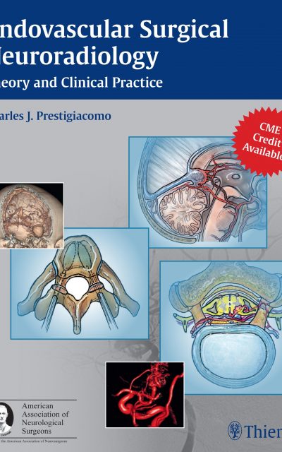
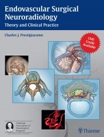
 Editor: Charles J. Prestigiacomo, MD
Contributing Editors: E. Jesus Duffis, MD; and Chirag D. Gandhi, MD
Publisher: Thieme – 722 pages with 716 illustrations
Book Review by: Nano Khilnani
The editors point out in the Preface of this work that explosive growth of knowledge, from the late 1920s onwards, is characteristic of the subspecialty field of endovascular surgical neuroradiology. What is significant for that period is the success that Egas Moniz, a Portuguese neurologist, encountered in cerebral angiography.
Moniz is regarded as one of the founders of what in earlier decades was referred to as “psychosurgery”. He developed the surgical procedure known then as leucotomy, and better known today as lobotomy. He was the first Portuguese national to receive a Nobel Prize. He shared that honor in 1949 with Walter Rudolf Hess, a Swiss physiologist.
Since then, due to the growth of technology and the bold and innovative thinking of pioneers in interventional medicine, people who had conditions that were inoperable, have been helped. Those conditions are now treatable and thousands of patients have benefitted from that technological growth and courageous innovation.
This textbook is useful to you the student, intern, resident or practitioner in this field because of the following:
- It brings into focus in a single resource the various current and up-to-date devices, procedures, and techniques used by top experts in their respective specialties.
- It also provides to you the benefit of their knowledge, experience, and insight, which guide you to avoid complications, or if they by chance occur, complication management.
- It is comprehensive and exhaustive, yet practical and usable. It reflects knowledge obtained through research as well as practical solutions to patients’ various problems.
One hundred and forty experts from all over the United States contributed material for this textbook that is presented in 59 chapters in this book, and organized in eleven parts. They are physicians and surgeons (and some nurses) specializing in cerebrovascular medicine and surgery, children’s medicine, endovascular medicine and surgery, interventional neuroradiology, neurology, neuroradiology, neuroscience, neurosurgery, radiology, skull base surgery, surgical angiography, and related fields.
We present to you here below the titles of the eleven parts of this reference and practice book to give you an overview of the subjects covered in it:
- General Endovascular Tenets
- Perioperative Care
- Angiographic Fundamentals
- Intracranial Aneurysms and Vasospasm
- Arteriovenous Malformations
- Dural Arteriovenous Malformations
- Lesions of the Head and Neck
- Atherosclerotic Disease
- Acute Stroke Interventions
- Lesions of the Spine
- Additional Endovascular Applications
This book has three main objectives:
- Explain the angiographic anatomy, physiology and pathophysiology of cerebrovascular disease as it pertains to acute ischemic stroke, aneurysms, arteriovenous malformations, atherosclerotic disease, and tumors.
- Describe the treatment options for acute ischemic stroke, aneurysms, arteriovenous malformations, and atherosclerotic disease, and apply them in clinical practice.
- Recognize the potential pitfalls for all endovascular therapeutic options and apply the learned principles for the avoidance and/or management of the complications.
Organization and presentation of material in the chapter is smart and useful. In the first chapter entitled Vascular Biology of the Endothelium for example, the authors Gaurav Gupta and Charles Prestigiacomo present knowledge and discussions under topics entitled:
- History
- Embryology
- Endothelium and the Subendothelial Matrix
In this chapter, Table 1.1 presents three Functions of tight junctions of the blood-brain barrier:
- Regulation of transmembrane tight junction proteins via G proteins, tyrosine kinases
- Regulation of the intracellular and extracellular calcium and cyclic adenosine monophosphate (cAMP) levels
- Regulation of intracellular metabolism via proteases and tumor necrosis factor alpha (TNFR-a)
After a discussion on the blood-brain barrier, Figure 1.2 shows two color sketches. The top one is a drawing of a blood vessel with an intact endothelium; and the lower one depicts a blood vessel with a damaged endothelium.
Detailed discussions and graphics are some of the notable features of this one-of-a-kind book. A Summary is provided in the chapter to help you remember the main points of the chapter, followed by a large list of References, to give you the opportunity to broaden your knowledge of the subject of the chapter.
Editor:
Charles J. Prestidigiacomo, MD, FAANS, FACS is Professor and Chairman of the Department of Neurological Surgery; Professor of Radiology, Neurology, and Neuroscience, and Director of Cerebrovascular and Endovascular Surgery at Rutgers New Jersey Medical School in Newark, New Jersey.
Contributing Editors:
E. Jesus Duffis, MD is involved with Neurointerventional Surgery in the Department of Neurology at Baystate Medical Center in Springfield, Massachusetts.
Chirag D. Gandhi, MD, FACS, FAANS is Associate Professor of Neurological Surgery, Neurology, and Neuroscience in the Department of Neurosurgery at Rutgers New Jersey Medical School in Newark, New Jersey.

