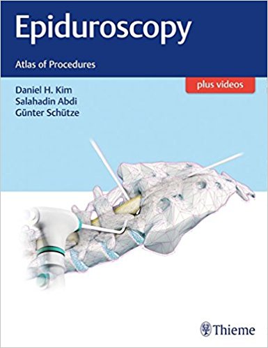
 Editors: Daniel H. Kim, MD; Salahadin Abdi, MD; and Gunter Schutze, Dr.med
Publisher: Thieme – 245 pages, with 360 illustrations
Book Review by: Nano Khilnani
Epiduroscopy is essentially the endoscopy of the spinal canal. It consists of inserting a fiber-optic scope into the narrow epidural space of the spine to diagnose and treat chronic back pain and various other spine conditions..
Different instruments have been developed over the years since the 1931 for examination of the spine, as discussed in the first chapter of this book – A History of Epiduroscopy. Michael Burman is cited as the first person to describe the visualization of the spinal canal and what he saw in there. He was an orthopedic surgeon working at the Hospital for Joint Diseases in New York. But it was a rigid arthroscopic instrument that he developed, compared to today’s flexible scope.
Five years later, in 1936, Elias Stern from Columbia University developed what was called a ‘spinascope’ which was used to look at the contents of the spinal canal after administering anesthesia. About a year later, in 1937, J. Lawrence Pool, an American neurosurgeon, used a myeloscope to improve visualization and diagnosis of patients with lumbar-sciatic syndrome anesthesia. His initial challenge was bleeding which obscured his view. Fortunately, Pool improved his technique and was able to visualize and report about the cauda equina syndrome (CES) and blood vessels.
Thirty-five specialists in anesthesiology, pain medicine, neurosurgery, orthopedic and spine surgery – mainly from the United States and Korea, as well as from Germany, Portugal, and Taiwan – authored the 27 chapters of this book focused on locating the sources of pain and treating it. The titles of this highly illustrated, valuable text are the following:
- The History of Epiduroscopy
- Applied and Radiological Anatomy of the Epidural Space for Spinal Endoscopy
- Pathophysiology of Spinal Pain and Pain Pathways
- Anatomic Origin of Spinal Pain and Clinical Manifestations
- Diagnostic Studies and Radiographic Imaging Modalities for Spine-Related Pain
- Epiduroscopy Equipment
- Epiduroscopic Surgical Tools
- Clinical Indications and Considerations for Epiduroscopy
- Sacral Hiatus Epiduroscopic Approach
- Transforaminal and Interfaminar Approaches in Epiduroscopy
- Epidusoscopic Images of Spinal Anatomy
- Evaluation of Epidural Pathology and Epiduroscopic Images
- Diagnostic and Functional Epiduroscopic Procedures
- Epiduroscopic, Histopathologic, and Microbiological Findings
- Anesthetic Management of Epiduroscopy
- Step-by-Step Epiduroscopic Techniques
- Spinal Epiduroscopic Mechanical Adhesiolysis
- Spinal Epiduroscopic Laser Therapy
- Epiduroscopic Procedure Using the 1,414 mm Nd:YAG Laser (Lutronic Laser)
- Spinal Epiduroscopic Radiofrequency Therapy
- Transforaminal Epiduroscopic Laser Annuloplasty for Diskogenic Pain
- Spinal / Epidural Analgesic and Pharmacologic Therapy
- Epiduroscopy-Assisted Spinal Cord Stimulator Electrode Implantation
- Epiduroscopic Ozone Therapy
- Patient Assessment for Epiduroscopy
- Epiduroscopy: Complications and Complication Avoidance
- Clinical Outcomes of Epiduroscopy
You can view 46 videos on epiduroscopy at www.MediaCenter.Thieme.com
While in the past, epiduroscopy had lagged behind open surgery in the diagnosis and management of spine disorders, its technology has now advanced ahead in recent years. So those who use it now can access, visualize, and treat spine pathologies, the editors point out in the Preface.
Another reason why epiduroscopy of late has become a widely used diagnostic tool is explained by the editors: “Since many of the spine pathologic clinical symptoms result from the interaction of neural elements and its surrounding structures in the epidural space, epiduroscopy provides a means for the diagnosis and treatment through direct visualization and focused intervention.”
Indeed, this is another example of the beneficial effects of the onward march of technology to solve problems and make our better. Long live epiduroscopy! Until something better comes along that will improve on it?
This is a good book by a capable team, on a new means to look inside the spine and make it function better. Thieme is ever at the forefront looking for ways to enhance and improve your medical practice !
Editors:
Daniel H. Kim, MD is the Nancy, Clive, and Pierce Runnells Distinguished Chair in Neuroscience, Professor and Director of Spinal Neurosurgery, Reconstructive Peripheral Nerve Surgery, Director of the Microsurgical Robotic Lab in the Department of Neurosurgery at the University of Texas
Salahadin Abdi, MD is Professor and Chairman of the Department of Pain Medicine, the Helen Buchanan and Stanley Joseph Seeger Endowed Research Professor at the University of Texas MD Anderson Cancer Center in Houston, Texas,
Gunter Schutz, Dr.med is Director of Anesthesiology, Critical Care Medicine, and Pain Medicine at Marien Hospital Iserlohn Privatpraxis fur Spezielle Schemerztherapie in Dortmund, Germany.


