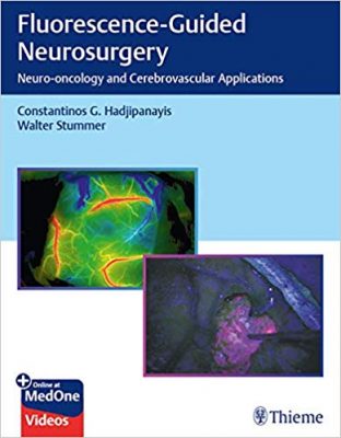
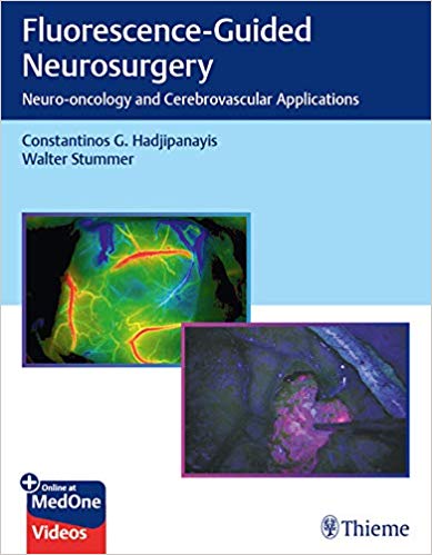
 Editors: Constantinos G. Hadjipanayis MD and Walter Stummer MD
Publisher: Thieme – 149 pages, with 74 illustrations
Book Review by: Nano Khilnani
Fluorescence-guided surgery (FGS) is a step forward for better illumination and visualization in determining which structures are normal and which are abnormal. FGS is a technological advancement in medicine, providing better views than the current instruments used by neurosurgeons – the operative microscope and the endoscope, write the editors of this book – Dr. Hadjipanayis and Dr. Stummer.
In 1910, the neuro-endoscope was introduced in Dr. Victor Lespinasse, and Dr. Theodore Kurze introduced the operative microscope for neurosurgery in 1967.
In the early 1990s, a third method of better observation and localization – frameless stereotactic neuronavigation – was introduced. In medicine and surgery there is a constant need for better outcomes.
This book introduces and details this relatively new method of observation for neurosurgeons to pinpoint the areas where surgical removal of tumors is required. One of the editors of this book – Dr. Walter Stummer – described in 1998 for the first time the use of 5-aminolevulinic acid (5-ALA) in FGS of high-grade glioma (HGG) tumors in patients with the use of a modified operative microscope.
In 2003, Dr. Andre Raabe first describe the use of the fluorescent agent indocyanine green (ICG) to visualize the flow of blood in vessels exposed in surgery. This technique known as ICG videoangiography would be used by neurosurgeons during aneurysm surgery for confirmation of proper surgical clip ligation.
In 2007, the European Medicines Agency (EMA) approved use of 5-ALA for resection of HGG tumors. This major Europe-wide event heralded the birth of fluorescence-guided neurosurgery globally
In 2017, the Food and Drug Administration (FDA) in the United States approved use of 5-ALA Gleolan as an imaging agent to better detect in real time, malignant tissue during surgery on gliomas.
Fifty-nine specialists mainly in neurosurgery, but also in oncological sciences and in otolaryngology from all over the United States and five other countries – Austria, Germany, Japan, Spain, Switzerland – authored the 20 chapters of this book. We show them below as an overview of the contents of this book:
- Current Fluorescence-Guided Neurosurgery and Moving Forward
- Designing and Reporting Studies on Intra-operative Tissue Imaging in the Brain
- 5-Aminolevulinic Acid and High-Grade Gliomas
- 5-Aminolevulinic Acid and Recurrent High-Grade Gliomas
- 5-Aminolevulinic Acid and Recurrent Low-Grade Gliomas
- Intra-operative Fluorescence Guidance in Meningiomas
- 5-Aminolevulinic Acid and Brain Metastases
- 5-Aminolevulinic Acid and Indocyanine Green: Fluorescence-Guided Resection of Spinal Cord Intramedullary Tumors
- 5-Aminolevulinic Acid in Pediatric Brain Tumors, Other Adult Tumors, and Photodynamic Therapy
- Fluorescein-Guided Tumor Resection in Neurosurgical Oncology
- Fluorescein and High-Grade Gliomas
- Second Window Indocyanine Green: Near-Infrared Optical Contrast for Intraoperative Identification of Brain Tumors
- Cancer-Targeted Alkylphosphocholine Analogs for Intraoperative Visualization
- Tozuleristide Fluorescence-Guided Surgery of Brain Tumors
- Confocal Endomicroscopy
- Fluorescence-Guided Surgery, Intra-operative Imaging, and Brain Mapping (MIR, DTI, and Cortical Mapping
- Raman Spectroscopy and Brain Tumors
- Indocyanine Green and Cerebral Aneurysms
- Indocyanine Green Videoangiography and Arteriovenous Malformations
- Indocyanine Green and Cerebral Revascularization
This book essentially presents and describes all the current applications in fluorescence-guided neurosurgery, both in neuro-oncology and in cerebrovasacular surgery. It also presents a critical analysis of intra-operative tissue imaging.
These are the potential advantages and disadvantages of intra-operative optical tissue imaging:
Advantages:
- Real-time
- Accustomed magnification
- Brain shift is no concern
- Full integration into the surgical microscope
Disadvantages:
- To some extent, loss of normal optical information while using the optical imaging strategy
- Time dependency of selected methods
- Regulatory issues regarding drugs and devices
- Two-dimensional representation
- Signal often obscured during surgery (blood, hemostatic agents)
- Signal alteration by surgical manipulation
This book has an excellent presentation of a pioneering technique in better visualization and location of tumors: fluorescence-guided neurosurgery.
Editors:
Constantinos G. Hadjipamayis MD, PhD is Professor and Site Chair, Department of Neurosurgery at Mount Sinai Downtown Union Square /Beth Israel; Professor of Oncological Sciences at Icahn School of Medicine at Mount Sinai, and Director of Neurological Surgery at Mount Sinai Health System in New York, New York
Walter Stummer MD, PhD is Professor and Chairman of the Department of Neurosurgery at the University of Munster in Munster, Germany.


