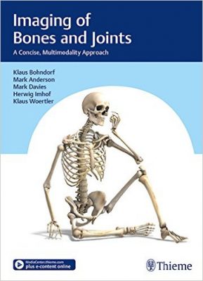
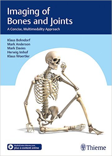
 Editors: Klaus Bohndorf, MD; Mark W. Anderson, MD; Mark Davies, MRCP; Herwig Imhof, MD; and Klaus Woertler, MD.
Publisher: Thieme – 513 pages, with 2,181 illustrations
Book Review by: Nano Khilnani
This is a comprehensive yet concise text on musculoskeletal imaging. It covers different types of disorders found in various parts of the human skeletal system by using a number of imaging modalities such as computed tomography (CT), magnetic resonance (MR), radiography, ultrasound equipment, and other means.
The disorders covered in this book are of various types such as infections, lesions, tumors, or others. The abnormalities shown and discussed within this book are congenital, hormonal, metabolic, rheumatic, toxic, or of a different nature.
Thirty-five specialists mainly from Germany, and four other countries – Austria, Switzerland, the United Kingdom, and the United States authored or coauthored the content in this book on imaging of bones and joints.
They are mainly biomedical imaging specialists, image-guided therapists, radiologists, and orthopedic surgeons teaching at universities and / or working in hospitals. To give you an overview of the coverage of this book, we provide you below the names of its chapters:
- Acute Trauma and Overuse Injuries
- Acute Trauma and Chronic Overuse
- Infections of the Bones, Joints, and Soft Tissues
- Tumors and Tumor-like Lesions of Bone Joints, and the Soft Tissues
- Bone Marrow
- Osteonecroses of the Skeletal System
- Osteochondroses
- Metabolic, Hormonal, and Toxic Bone Disorders
- Congenital Disorders of Bone and Joint Development
- Rheumatic Disorders
- Miscellaneous Bone, Joint, and Soft Tissue Disorders
- Interventions Involving the Bone, Soft Tissues, and Joints
This work can be used in several ways:
- As an overview of the essentials of musculoskeletal imaging
- As an aid in getting started in the medical specialty of bone and joint radiology
- As a reference work for study and medical practice
- As a faithful guide right up to and beyond board exams
The content in this book is organized around clinical disorders. It describes the findings of all modalities relevant to each disorder in its chapters. To get more detailed information on a given area, we recommended that you obtain subspecialty textbooks.
This is a book that is “cast from a single mold,” a description that is sometimes used to describe a unified style of presentation of different and varied materials. The editors have revised, trimmed, supplemented, and harmonized text and images so they become a unified whole.
The editors write that comprehensive books on a given specialty such as this one, are “an ideal tool for gaining an overview, learning the basic principles, and acquiring knowledge in the field of musculoskeletal radiology.”
One of the outstanding values of this book is the large number of images used: 2,181 or an average of more than 4 images per image. These greatly help in viewing and analyzing the conditions and diseases within the bones, in the bone marrow, in the joints, in regular and soft tissue, and in other places. For example, page 7 shows 12 types of fractures.
The range of topics presented and discussed in any given chapter is fairly extensive. In chapter 8 entitled Metabolic, Hormonal, and Toxic Bone Disorders for example, you will find the following topics and subtopics discussed along with images showing the conditions.
8.1 Osteoporosis
8.1.1 Classification and Clinical Presentation f Osteoporosis
8.1.2 Bone Density Testing
8.1.3 Radiographic Findings in Osteoporosis
8.2 Rickets and Osteomalacia
8.3 Hyperparathyroidism and Hypoparathyroidism
8.3.1 Hyperparathyroidism
8.3.2 Hypoparathyroidism
8.4 Renal Osteodystrophy
8.5 Drug-Induced Changes to the Bone
8.5.1 Corticosteroids
8.5.2 Other Drugs
8.6 Amyloidosis
8.7 Other Osteopathic Diseases
8.7.1 Hemophilic Arthopathy
8.7.2 Acromegaly
This book covers many of the clinical disorders pertaining to bones, joints, and surrounding tissue, and provides you a huge number of images obtained through several modalities, to enable you to see and analyze them.
In summary, it provides an overview, gets you started in the medical specialty of radiology, serves as a reference manual, is a reliable guide to help you prepare for the board exams, and is a keepsake for well beyond that milestone.
As a plus, the contents of this book, and much more, can be viewed online by going to: www.MedicalCenter.Thieme.com and registering your copy, by inputting the code shown on the inside front cover of the book.
Editors:
Klaus Bohndorf, MD is a Professor of Radiology in the Department of Biomedical Imaging and Image-Guided Therapy at the Medical University of Vienna, in Vienna, Austria.
Mark W. Anderson, MD is a Professor of Radiology in the Department of Radiology at the University of Virginia in Charlottesville, Virginia.
Mark Davies, MRCP, FRCR is Professor of Radiology in the Department of Radiology at Royal Orthopedic Hospital in Birmingham, UK.
Herwig Imhof, MD is Professor of Radiology in the formerly Department of Biomedical Imaging and Image-Guided Therapy at the Medical University of Vienna, in Vienna, Austria.
Klaus Woertler, MD is Professor of Radiology in the Department of Radiology at the Technical University of Munich in Munich, Germany.


