 Editors: Haresh Kirpalani, BM; Monica Epelman, MD; and John Richard Mernagh, MD
Publisher: Cambridge University Press – 257 pages
Book Review by: Nano Khilnani
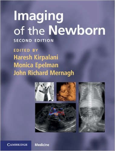
The two main purposes of this book according to the editors in their Preface are: first, that the members of neonatology healthcare team in the care of the newborn infant “speak the same language” as the radiology team with regards to presentations, differential diagnoses, and the like. And secondly, in the face of enormous learning demands on the concerned healthcare teams, there should be a strong emphasis and focus on a single, most important aspect of the clinical assessment of the infant.
Twenty-five specialists in neonatology, nephrology, obstetrics, pediatrics, radiology, and related fields, from around the United States and three other countries – Australia, Canada, and the United Kingdom – authored the 13 chapters of this compact book. We list its chapters below to give you overview of its contents:
- Introduction to principles of the radiological investigation of the neonate.
- Evidence-based use of diagnostic imaging: reliability and validity
- The chest
- Neonatal congenital heart disease
- Special considerations for neonatal extracorporeal membrane oxygenation (ECMO)
- The central nervous system
- The gastrointestinal tract
- The kidney
- Some principles of in-utero and post-natal formation of the skeleton
- Metabolic diseases
- Catheters and tubes
- Routine prenatal screening during pregnancy
- Antenatal diagnosis of selected defects
This book is ideal for pediatric and junior radiology residents, clinicians and others involved in neonatology, physician assistants, and obstetric and pediatric nurses.
Among the features new to this edition are the following:
- Images updated throughout to reflect improvements in equipment and scanning techniques
- Expanded chapters on cardiovascular problems, bone, and prenatal ultrasound
- New chapters on clinical utility of procedures, ECMO, metabolic and inborn errors
of metabolism, and antennal diagnosis of common abnormalities
“Imaging in newborns remains a great intellectual challenge,” writes Dr. Alan Daneman in his Foreword to this book. While plain radiographs can and do reveal conditions of many neonates that can be appropriately managed, there are some conditions and situations that can only be adequately viewed through more sophisticated imaging modalities. Here are some that he mentions:
- Computed tomography
- Contrast examinations of the gastrointestinal tract
- Interventional procedures
- Magnetic resonance imaging
Furthermore, “meticulous technique” is required in examining these very small patients. In the newborn, Dr. Daneman writes, special attention must be given to such considerations as:
- Immobilization techniques
- Temperature control
- Ventilation during imaging
Imaging results of the newborn are insufficient when viewed without clinical history of the patient recorded by the doctor charged with his or her care and/or neonatal surgeon. Needless to say, close consultation and ongoing dialogue with all involved persons is important for the best outcome to be achieved.
Let us take a look inside at chapter 4, Neonatal Congenital Heart Disease written by Drs. Jeffrey C. Hellinger and Mary M.K. Seshia to give you an idea of the structure of the contents you will find in other chapters.
Like all other chapters, a lot of images of various kinds with detailed captions, are presented. In the Introduction to this chapter, the authors state that several imaging means are used to view the chest are of the infant to examine congenital heart disease (CHD).
Among these are: catheter angiography, computed tomographic angiography (CTA), magnetic resonance angiography (MRA), magnetic resonance imaging (MRI), radiographs, and ultrasound (echocardiography, echo).
The first image is a radiograph of a child with tricuspid atresia. The second image shows how an MRA-MRI was used to diagnose a double aortic arch in a patient presented with stridor. A third image shows the use of CTA to diagnose iatronic aortic dissection in a patient with Ehlers-Danlos syndrome, recovering from tetralogy of Fallot repair. A fourth image shows a vascular ring diagnosed non-invasively and without radiation, using MRA.
What follows in this chapter are descriptions and the details on the use and outcomes of each of the imaging modalities we mention above, when examining infants with CHD. The basic headings in this chapter are the following:
- Introduction
- Imaging Modalities
- Cardiovascular MRI and CT
- Modality Interpretation: A Systematic Approach
- Clinical Diagnosis of Congenital Heart Disease
- References
There’s much to be learned in this chapter and the rest of this book. It’s a handy guide that’s packed with a lot of important and even essential information expertly laid out using text, images, charts, drawings, tables, and other visuals. A very valuable book, and I’ll say a must for any aspiring as well as practicing neonatologist, or any professional involved in the care of infants.
Editors:
Haresh Kirpalani, BM, MRCP, FRCP (UK), FRCP (C) MSc (Clinical Epidemiology) is Professor of Pediatrics at University of Pennsylvania, Division of Neonatology, The Children’s Hospital of Philadelphia, in Philadelphia, Pennsylvania, USA. He is also an Associate Member of the Department of Clinical Epidemiology and Biostatics at McMaster University in Hamilton, Ontario, Canada.
Monica Epelman, MD is Assistant Professor of Radiology at the University of Pennsylvania, and Director of Neonatal Imaging at The Children’s Hospital of Philadelphia in Philadelphia, Pennsylvania, USA.
John Richard Mernagh, MD, PhD, FRCPC is Associate Professor of Radiology at McMaster University Medical Centre, and McMaster Children’s Hospital in Hamilton, Ontario, Canada.


