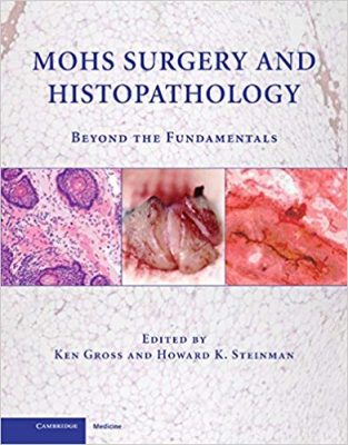
 Editors: Ken Gross, MD and Howard K.Steinman, MD
Publisher: Cambridge University Press – 184 pages
Book Review by: Nano Khilnani
Mohs surgery, developed in 1938 by an American general surgeon named Frederic E. Mohs, is a precise surgical technique used to treat skin cancer. During Mohs surgery, thin layers of cancer-containing skin are progressively removed and examined until only cancer-free tissue remains. Mohs surgery is also known as Mohs micrographic surgery
According to a Wikipedia article, the cure rate with Mohs surgery cited by most studies is between 97 and 99.8 percent for primary basal-cell carcinoma, the most common type of skin cancer. Mohs procedure is also used for squamous cell carcinoma, but with a lower cure rate.
Recurrent basal-cell cancer has a lower cure rate with Mohs surgery, more in the range of 94 percent. It has been used in the removal of melanoma-in-situ (cure rate 77 to 98 percent depending on surgeon) and certain types of melanoma (cure rate 52 percent).
Note that cancer that begins in squamous cells is known as squamous cell carcinoma. These cells are thin, flat cells that look like fish scales, and are found in the tissue that forms the surface of the skin, the lining of the hollow organs of the body, and the lining of the respiratory and digestive tracts.
Twenty specialists (including the two editors above) in dermatologic and skin cancer surgery, otorhinoaryngology, and plastic and reconstructive surgery wrote the chapters of this book. All are from the United States except one – Ofer Arnon – who is from Israel. Here below are the 20 chapters of this book, organized around four Parts:
- Part I – Microscopy and Tissue Preparation
- Introduction
- How to Excise Tissue for Optimal Sectioning
- Optimizing the Mohs
- Tissue preparation and Chroma-coding
- Embedding the Techniques
- Part II – Introduction to Laboratory Techniques
- Lab Pearls: Making Great Slides
- Lab Pearls: Staining, Inking and Cover-slipping
- Lab Pearls: Troubleshooting Slide Quality
- MOHS Slides Organization and Standardization for Effective Interpretation
- MOHS Mapping
- Part III – Microanatomy and Neoplastic Disease
- Normal Micro-anatomy: Vertical and Horizontal
- Baal Cell Carcinoma: Vertical and Horizontal
- Squamous Cell Carcinoma: Vertical and Horizontal
- Unusual Tumors: Vertical and Horizontal
- MOHS for Melanoma
- Taking Stages Beyond Stage I
- Perineural Tumors
- Part IV – Special Techniques and Stains
- Fixed-Tissue Mohs
- Toluidine Blue Stain for Mohs
- Forms and Templates for Mohs Surgery
Mohs surgery is known to be a highly effective technique for the removal of most types of cutaneous and oral pharyngeal cancers. The procedure used in this type of surgery allows for not only a precise, but also complete removal of cancers while maximizing the preservation of surrounding normal tissue.
In this book, images are shown of the tumor foci, and the precise surgical margins, and other relevant findings that can correlated with their locations on the surgical wound.
The editors – Drs. Ken Gross, MD and Howard K.Steinman – write at the beginning of this book:
“The ability to create perfect slides for histological examination lies at the core of Mohs surgery
They also note:
“The procedure has the highest cure rate among alternate cancer treatment modalities for the cancers for which it is utilized.”
In sum, this book essentially describes and illustrates the methods that the Mohs surgeon-pathologist and the Mohs lab technician utilize to optimize the Mohs technique and produce the highest-quality slides and highest cure rates possible, and it breaks new ground in describing techniques.
Editors:
Ken Gross, MD is a non-salaried Clinical Professor in the Division of Dermatology at the University of California School of Medicine in San Diego. He is also in private practice limited to the treatment of skin cancer in San Diego, California.
Howard K. Steinman, MD is Director of Dermatologic and Skin Cancer Surgery and Associate Professor of Dermatology at the Texas A&M University Health Sciences Center College of Medicine, Scott and White Medical Center in Temple, Texas
Contributors:
Ofer Arnon, MD, Norman A. Brooks, MD, John B. Campbell, MD, Laura T. Cepeda, MD,
A Neil Crowson, MD, Carlos Garcia, MD, Leonard H. Goldberg, MD, Ken Gross, MD, Arash Kimyai-Asadi, MD, Alex Lutz, MD, Adam J. Mamelak, MD, Alexander Miller, MD, Tri H. Nguyen, MD, Terence O’Grady, MD, Michael Shelton, MD, Daniel M. Siegel, MD, Howard K. Steinman, MD, and Edward H. Yob, DO.


