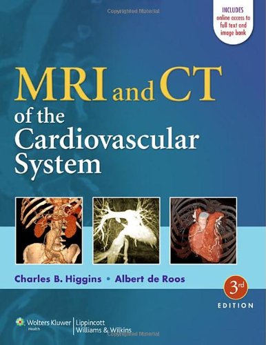
 Editors: Charles B. Higgins, MD; and Albert de Roos, MD
Publisher: Wolters Kluwer | Lippincott Williams & Wilkins – 676 pages
Book Review by: Nano Khilnani
This book is about effectively and thoroughly using two relatively recent scanning and imaging technologies in diagnosing cardiovascular conditions and taking the necessary further steps in treating patients.
The authors point out that these technologies covered in this book are relatively new, but their effectiveness in diagnosing heart disease has advanced rapidly in recent years. Computed tomography or CT produced the first images of the beating heart in the late 1970s, and the first images of the heart using magnetic resonance or MR were obtained around 1982.
Sixty-eight specialists – in biomedical engineering, cardiology, internal medicine, nuclear medicine, oncology, physiology, radiology, vascular surgery, and allied fields – from the United States and Belgium, Canada, France, Germany, Italy, Japan, The Netherlands, Spain, Sweden, Switzerland, and the United Kingdom authored or contributed to the 39 chapters of this book.
All these chapters – covering a wide range of imaging techniques – have been organized around six Parts. To give you are overview of the material you will find in special book on CT and MR, they are:
- Basic Principles
- Clinical Approach to Cardiovascular Magnetic Resonance Techniques
- Magnetic resonance Angiography Techniques
- Techniques for Cardiovascular Computed Tomography
- Cardiac Anatomy and Physiology: Imaging Aspects
- Blood Flow Measurements
- Quantification in Cardiac MRI and CT
- CVMR: Imaging Planes and Anatomy
- Coronary CTA: Display Techniques and Planes
- Cardiovascular MR in Infants and Children
- Acquired Heart Disease
- Magnetic Resonance of Cardiomyopathies and Myocarditis
- Arrhythmogenic Right Ventricular Cardiomyopathy / Dysplasia
- MRI and CT of Pericardial Diseases
- Cardiac and Pericardiac Masses
- Valvular Heart Disease
- Ischemic Heart Disease
- Myocardial Perfusion in Ischemic Heart Disease
- Assessment of Ischemic Heart Disease Utilizing Dobutamine
- Assessment of Myocardial Viability by Contrast Enhancement
- Delayed Enhancement in Nonischemic Myocardial Disease
- Magnetic Resonance Imaging Risk Assessment in Ischemic Heart Disease
- Coronary Magnetic Resonance Angiography: Technical Approaches
- Coronary Artery Imaging – Clinical Approaches
- Coronary Blood Flow Measurements
- Cardiovascular MR and CT of Coronary Artery Bypass Grafts
- CT Imaging of Coronary Artery Calcification
- Coronary CT Angiography
- CT Evaluation of Myocardial Perfusion. Function, and Late Enhancement
- Atherosclerotic Plaque Imaging: MRI
- Plaque Characterization: CT
- Congenital Heart Disease
- Congenital Heart Disease: MR Evaluation of Morphology And Function
- MR Imaging of Function and Flow in Postoperative Congenital Heart Disease
- Adult Congenital Heart Disease
- Vascular Diseases
- MRI and Ct of Thoracic Area
- Thoracic Aorta Disease: Flow Evaluation by MR
- CT Angiography of the Abdominal Arteries
- MR and CT Angiography of Peripheral Arteries
- Interventional MRI
- Catheter Tracking and Devices
- Endovascular Interventional MRI
- Endovascular Interventions – Congenital Heart Disease
- Endovascular Delivery of Gene and Stem Cell Therapy
This book comes with access to the complete contents online, fully searchable, plus other valuable features. Simply do the following to gain instant access and register your book:
- Visit http://silution.lww.com
- Enter access code by scratching out grey tab found on the inside front cover of your book
- Follow the instructions to activate your access
The website named above is for individual use only. If you are interested in multi-user access, call customer service at 1-800-468-1128 in the United States or 1-410-528-4000 outside the U.S.
Or email: customerservice@lww.com.
Systematic organization of the vast amount of materials including data is a notable hallmark of this valuable book focused on the comprehensive use of MR and CT techniques in scanning and imaging patients to determine disease in their hearts and blood vessels.
An Introduction – a paragraph or a few of them – is provided in the beginning to give you an overview of each chapter’s contents. This tops the bulleted points. It is followed by various topics.
For example in chapter 1 entitled Clinical Approach to Cardiovascular Magnetic Resonance Techniques, the topics that follow the introductory text are: Coils, Cardiac Motion Compensation, Respiratory Motion Compensation, Survey, Plan Scan, Anatomy, Function, Perfusion, Delayed Enhancement, Flow, Coronary MRA, Vessel Wall Imaging, CVMR Image Processing, Reduced Data Acquisition Methods, and Appendix: Calculations on K-Space.
Numerous CT and MR scans (mostly black and white, but some with color), charts, diagrams, tables and other forms of visual presentation are used in each chapter to convey information and analysis to you.
A Summary concluding the basic points of each chapter helps you retain its important highlights. The Acknowledgements section thanks those who contributed information to that chapter. And typically, long lists of References are provided to you at the end of each chapter for your further study of relevant topics.
This is an excellent resource on the use of CT and MR imaging technologies as it pertains to the cardiovascular system, especially when you consider all the additional information available online. The doctors named below have really done a marvelous job in this third edition published in 2013.
Charles B. Higgins, MD is Professor of Radiology at the University of California in San Francisco, California.
Albert de Roos, MD is Professor of Radiology in the Department of Radiology at Leiden University Medical Center in Leiden, The Netherlands.


