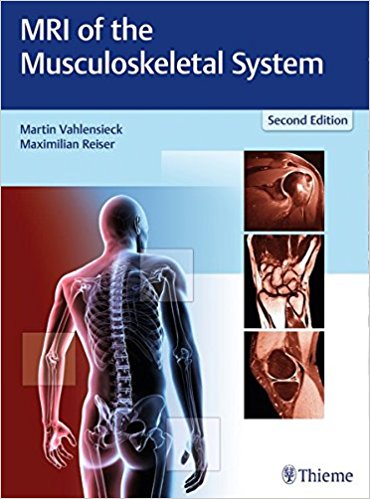
 Editors: Martin Vahlensieck, MD and Maximilian Reiser, MD
Publisher: Thieme – 677 pages, with 2,062 illustrations
Book Review by: Nano Khilnani
The musculoskeletal system is quite pervasive, in that it is part of almost the entire human body.
From a layman’s viewpoint, the human skeleton is the frame upon which our whole body is built, and bones are found within even small areas, such as our ears. And our muscular system is firmly based on our skeletal frame.
This book deals with magnetic resonance imaging or MRI of the various major parts of our musculoskeletal system. It is a large-size (9”x12”), massive (677 pages), and updated textbook that contains 2,062 images covering the entire musculoskeletal system of the body.
With an average of over three images per page with detailed captions, it is an important resource to own and have at hand as a reliable ready-reference manual, not only for you if you are an established radiologist or training to be one, but also if you are a specialist on any area of the human anatomy.
Twenty-two clinical, interventional, and musculoskeletal radiologists, mainly from Germany, but also some from Austria, Switzerland, and the United States, authored the 15 chapters and Appendix of this book that we name below.
This second edition released in 2018 contains numerous updates and additions to the first edition that was released 10 years ago in 1999. But before we list those updates, let us take a look at an overview of what you will find in it – a list of the chapters:
- Relevant Magnetic Resonance Imaging Techniques
- The Spine
- The Shoulder
- The Elbow
- The Wrist and Fingers
- The Hip and Pelvis
- The Knee
- The Lower Leg, Ankle, and Foot
- The Temporomandibular Joint
- The Muscles
- Bone Marrow
- Bone and Soft Tissue Tumors
- Osteoporosis
- The Sacroiliac Joints
- The Jaws and Periodontal Apparatus
- Appendix – Other Disorders and Diagnoses
Among the updates and new content you will find in this second (2018) edition are:
- Images that were taken with open high-field systems that have advantages for claustrophobic patients
- Superior Images obtained with 3 Tesla scanners
- Images that relate to sequence acquisition
- Images that are reflective of technical innovations such as MRI neurography and MRI prosthesis sequences
- Continually more accurate imaging processes that have replaced older classification systems. Example: meniscus diagnostic imaging is no longer used to characterize increases in signal intensity but rather to classify ruptures, in terms of the implied type of tear and its extension, or to detect even the most minute ligament injuries, for example, of the fingers.
- Introduction of a myriad new graphics and reference sources, replacement, and addition of several illustrations
- In many places, contributing authors have referred Internet sites deemed to be useful to readers who wish to obtain more information
- Research tips on more effective use of Internet search terms
- Sections on clinical relevance presented as Clinical Reviews where opinions of clinical radiologists are sought on the performance of the MRI
In sum, the major features of this book are the following:
- More than 2,000images of reference quality, the majority new for this edition
- Drawings, where helpful, aid the reader in identifying and delineating normal and pathological entities
- Includes all the latest advanced techniques: MT neurography and myelography, diffusion imaging, quantitative MRI, mDIXON, and more
- All MR exams described fully, with choice of sequence, positioning, choice of coils, when/how to use contrast, protocols
- Discussion of possible errors in interpretation
- Comparison of MR imaging with other modalities
- Tables expand and organize information on sequence parameters and differential diagnoses
Clear and accurate images of the musculoskeletal system enable the physician to look for and view disorders, and the extent of their spread and possible impact on the patient, Accurate and complete imaging is critical in determining the optimal treatment mode, including surgery, if needed. MRI, in comparison with other imaging modalities has proven to be among best.
The editors write that MRI “is the only imaging modality that enables visualization of all components of joints within single examination. Yet given the bewildering variety of possible sequence parameters, with and without contrast medium, acquiring and interpreting MR images with confidence is a challenge, requiring experience usually only gained after examining thousands of studies with a careful systematic approach.”
This comprehensive, well organized, authoritative, updated, and valuable book on MRI imaging of the musculoskeletal system is the key to making the right decision or recommendation for the best course of treatment for the patient.
Editors:
Martin Vahlensieck, MD is Professor of Radiology and in the practice of Radiology and Nuclear Medicine in Bonn, Germany.
Maximilian Reiser, MD, FACR, FRCR is Professor of Radiology, Chairman of the Department of Clinical Radiology, and Dean of Medicine at Ludwig Maximilian University in Munich, Germany.

