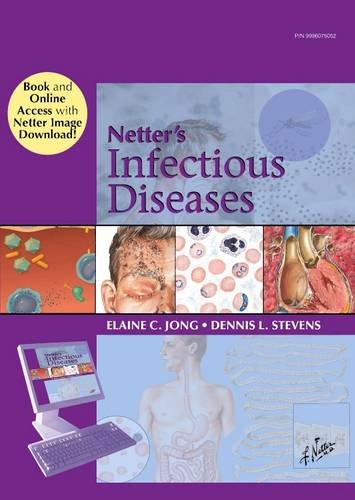
 Editors: Elaine C. Jong, MD; and Dennis L. Stevens, PhD, MD.
Illustrations by: Frank H Netter, MD
Publisher: Elsevier Saunders – 602 pages
Book Review by: Nano Khilnani
This book discusses in good detail, infections and the diseases caused by them, many of them shown in graphic form to enhance your understanding of them.
Ninety physicians and other specialists from all over the United States and seven other countries – Australia, Canada, Germany, Israel, Peru, South Africa, and Venezuela – authored the 93 chapters of this extensive book organized into nine sections, namely:
- Vaccine-Preventable Diseases in Children and Adolescents
- Skin and Soft-Tissue Infections
- Respiratory Tract Infections
- Systemic Infections
- Surgical Infections
- Sexually-Transmitted Infections
- Infections Associated with International Travel and Outdoor Activities
- Parasitic Diseases
- Emerging Infectious Diseases and Pandemics
You can access the contents of this print edition online by registering yourself as a user and registering this book as well:
User Registration
- Use the Register button at www.NetterReference.com
- Fill in all the required details; complete the registration by clicking on the Submit button
- Click on the link that comes up next, then follow steps in the Book Registration below
Book Registration
- You will be prompted to enter the PIN found by scratching off the gray sticker on the inside front cover of your book
- Once the book is registered, you will be taken to the book’s home page
- Access the book content by clicking on the book image
Detailed full-color depictions were the hallmark of all illustrations created by the late Dr Frank Netter during his lifetime. You will know they were drawn by him with his distinctive signature (reflecting great creativity, as surely a graphologist will tell you) shown on the right bottom of the images.
Where the drawings are of people, the illustrations are starkly life-like! For example, in chapter 2, Diphtheria and Tetanus, see the drawing (Figure 2-3: Generalized tetanus) on page 8 of the man (top right) who is having a spasm of the face, jaw, and neck muscles. This is sometimes called lockjaw. It is as if you are feeling the sudden, excruciating pain he must be feeling.
In the same sketch, the man is lying supine on the bottom right with his arms above the floor and the toes of his feet turned upward. The photo caption reads: “Complete titanic spasm in advanced disease. Patient rigid in moderate opisthotonos, with arms extended, abdomen boardlike. Respiratory arrest may occur.”
On this same illustration are five other drawings. On the top left drawing the caption reads: “Clostridium tetani, gram-positive spore-bearing rods.” This round image through a microscope shows you what this disease-causing organism looks like.
To the right of that top left drawing is one of a leg with a large wound, with the caption: “Organisms enter through large, small, or even unrecognized wound. Deep, infected punctures are most susceptible since organisms thrive best anaerobically.”
From the leg going southwest is a drawing of a nerve pointing to two other drawings: the first, of the spinal cord and the second, of the central nervous system, with the caption: “Toxin produced locally passes via bloodstream and lymphatics to peripheral nerve terminals and then travels along axons to central nervous system.”
Figure 2-3 is drawn in such detail and exquisite realism that it epitomizes the saying we often hear: “a picture is worth a thousand words.” What better way to learn than to see, and read the precise descriptions in the captions and accompanying text?
You will find numerous such illustrations throughout this book that will make learning the material so productive and so much fun. The systematic organization of material in the chapters also aids in facilitating the learning process for you.
You will find an Abstract at the beginning of each chapter, followed by discussions of the various topics (e.g. Risk Factors) with text broken up by boxes, charts, drawings, tables, and other items. At the end of each chapter you will find a box entitled Evidence followed by a list of references entitled Additional Resources.
Editors:
Elaine C. Jong, MD is Clinical Professor of Medicine Emeritus in the Divisions of Emergency Medicine and Allergy and Infectious Diseases at University of Washington School of Medicine in Seattle, Washington.
Dennis L. Stevens, PhD, MD is Chief of the Infectious Diseases Section at Veterans Affairs Medical Center in Boise, Idaho; and Professor of Medicine at University of Washington School of Medicine in Seattle, Washington.
Section Editors:
E. Patched Dellinger, MD
Thomas M. File, Jr., MD, Mac, MACP, FIDSA, FCCP
Jo Hoffman, MD
Jeanne M. Arezzo, MD, MPH
Chris Anna M. Mink, MD
Illustrations by:
Frank H Netter, MD. He was born in 1906 in New York City. He studied art at the Arts Students League and the National Academy of Design before entering medical school at New York University, where he received his MD degree in 1931. During his student years, his notebook sketches attracted the attention of the medical faculty and other physicians, allowing him to augment his income by illustrating articles and textbooks.
He continued illustrating as a sideline after establishing a surgical practice in 1933, but he ultimately opted to give up his practice in favor of a fulltime commitment to art. After service in the U.S. Army during World War II, Dr. Netter began in long collaboration with the CIBA Pharmaceutical Company (now Novartis Pharmaceuticals). The 45-year partnership resulted in the production of the extraordinary collaboration of medical art so familiar to physicians and other medical professionals worldwide.
Contributing Illustrators:
Carlos A.G. Machado, MD; John A. Craig, MD; James A. Perkins, MS, MFA; Tiffany S. DaVanzo, MA, CMI; and Anita Impagliazzo, MA, CMI.


