 Author: Duane E. Haines, PhD
Publisher: Wolters Kluwer | Lippincott Williams & Wilkins – 332 pages
Book Review by: Nano Khilnani
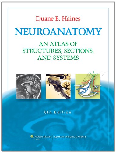
This latest edition of Neuroanatomy – An Atlas of Structures, Sections and Systems, while continuing to present detailed knowledge on the anatomy and functions, in the form of text and images on the human neurosystem, also introduces new information through computer tomography (CT) scans, magnetic resonance images (MRIs) and other visual means such as illustrations and actual black-and-white, single-color and full-color photos.
Put together and updated by Dr. Duane E. Haines – professor of neurology and neurosurgery at the University of Mississippi Medical Center – this newest edition with ten chapters also explains basic and clinical terms and concepts encountered in clinical settings; gives additional examples and presents new text; and provides a comprehensive overview of systems neurobiology.
The author emphasizes: “understanding systems neurobiology is the key element in the successful diagnosis of the neurologically compromised patient.”
There are six major changes in this updated edition. They are the following:
- Chapter 3 on cranial nerves has been revised and new clinical information has been added to it, with cross-references included to figures in other chapters.
- Examples of clinical conditions that have important anatomical correlates are demonstrated with a number of new images.
- In chapter 6, all black-and-white stained sections have been replaced with color versions, providing a high level of anatomical detail, particularly in the brain stem with many nuclei and tracts that have important clinical implications.
- Likewise in chapter 7, black-and-white versions of stained sections have been replaced with color images, enhancing clarity and visual impact of those images. Labeling has been changed to identify additional and important structures.
- Chapter 8 on neural systems has been revised and many improvements have been made, particularly the presentation of ten illustrations showing spinal and cranial nerve reflexes with accompanying descriptions.
- The questions and answers in chapter 10 have been revised and new ones added, reflecting the new information on basic and clinical science (with images included) in chapters 2 to 4 and chapter 8.
To give you a broad overview of the contents of this book produced on a large-page format, here’s what is covered in it:
- Introduction and Reader’s Guide
- External Morphology of the Central Nervous System
- Meninges, Cisterns, Ventricles and Related Hemorrhages
- Internal Morphology of the Brain in Unstained Slices and MRI
- Internal Morphology of the Spinal Cord and Brain in Stained Sections
- Internal Morphology of the Brain in Stained Sections: Axial-Sagittal Correlations with MRI
- Synopsis of Functional Components, Tracts, Pathways, and Systems: Examples in Anatomical and Clinical Orientation
- Anatomical-Clinical Correlations: Cerebral Angiogram, Magnetic Resonance Angiography and Magnetic Resonance Venography
- Q&As: A Sampling of Study and Review Questions, Many in the USMLE Style, All with Explained Answers
The Q&A section is very valuable. It contains 11 pages with more than 280 test questions to review – questions that the exam taker will encounter in the Subject National Board Examination and in Step 1 of the U.S. Medical Licensing Examination (USMLE).
The Index lists important words found in medical terminology texts and other entries that the student or doctor can look up, and go to the section in this book that contains the information on a specific topic. Color tabs on the top and on the right edges of the pages also make it easier to go to the chapter the reader is looking for.
Some of the other interesting and highly useful features of this excellent volume, besides the anatomical illustrations, CT scans, full-color photos, and MRI images, are pathway drawings of the nerves and review tables.
As in other books on health and medicine by this publisher, online resources are available to the purchaser on the contents of this book for study and review. An access code is needed, which is provided in a scratchable tab on the inside front cover.
Dr. Haines writes that there are two essential goals that an aspiring neurobiologist must attain: the first is to gain knowledge of this science and the relevant diagnostic skills to become competent in this field. The second is to pass the exams to acquire the necessary license to practice. The Q&A section helps you reach the second goal. And the book itself, organized and written very well by him, goes a long way in helping accomplish the first goal. We applaud his outstanding contribution to this field with this book.


