 Editor: Stephanie J. Doniger, MD
Publisher: Cambridge University Press – 357 pages
Book Review by: Nano Khilnani
Today there are numerous means of physically examining people beneath their skin to look for signs of disease or a disorder, and most of them require exposing the patient to radiation in various amounts. Some of these scanning methods are computed tomography (CT), computed axial tomography (CAT), and magnetic resonance (MR). There are numerous other imaging modalities as well, and depending upon the amount of radiation that our bodies are exposed to, they pose different levels of potential harm.
One of the earliest ways of looking inside our bodies followed the chance discovery of the x-ray in 1895 by Wilhelm Roentgen, a professor of physics in Worzburg, Bavaria (now part of Germany. He named his discovery ‘x-radiation’ because it was different from other types of electrical rays he was studying and he didn’t know what to call it. His later invention of an x-ray machine dramatically changed the history of medicine. With it, amazingly, the inner structures and later, functions of our bodies were made visible for the first time without cutting into flesh.
Many years earlier than the x-ray machine, beginning in 1816, doctors listened to what was going on inside our bodies with the stethoscope, which enabled them to listen to the heart and other organs, bringing into focus in their trained minds the images of those organs they had studied in their anatomy and physiology courses.
The ultrasound machine, which uses harmless sound waves instead of radiation to image the inside of the human body, was developed in the 1940s. The American acoustical physicist Floyd Firestone devised in 1940, the first ultrasonic echo imaging device, which he called the Supersonic Reflectoscope, to detect internal flaws in metal castings.
In 1941, the Austrian neurologist Karl Theo Dussik collaborated with his brother, Friedreich, a physicist, and the two were likely the first persons to ultrasonically echo image the human body, outlining the ventricles of a human brain. Ultrasonic energy was first applied to the human body for medical purposes by Dr. George Ludwig at the Naval Medical Research Institute in Bethesda, Maryland in the late 1940s.
As time went by, technological developments made it possible for ultrasound machines to become smaller, more powerful, and lighter, so they can be held in the hand. About four decades after the first ultrasound machine was developed, sonoscopes – sort of portable, mini ultrasound devices – made their way in the 1980s into emergency departments of medical centers in the United States.
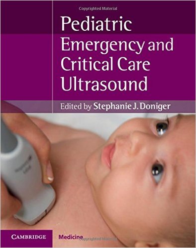
This book on the use of ultrasonography in pediatric emergency and critical care represents the work of its editor Dr. Stephanie Doniger and 32 other contributors from five countries – Canada, Italy, Saudi Arabia, the United Arab Emirates, and the United States – who authored or coauthored the 24 chapters of this book in four Sections that we name below:
- Section 1. Ultrasound fundamentals
- Introduction
- Physics and ‘knobology’
- Getting started
- Ultrasound in austere environments
- Section 2. Diagnostic ultrasound
- Introduction
- The extended focused assessment with sonography for trauma (E-FAST)
- Focused cardiac ultrasound
- Pulmonary ultrasound
- Inferior vena cava, aorta assessment
- HEENT (head, ears, eyes, nose, throat): ocular, sinus, neck ultrasound
- Renal, bladder
- The pediatric abdomen
- Hepatobiliary
- Male genitourinary, scrotal pain, swelling
- Pelvic obstetrics
- Pelvic gynecology
- Section 3. Procedural ultrasound
- Introduction
- Vascular access
- Bladder ultrasound for catheterization and supra-public aspiration
- Ultrasound-guided lumbar puncture
- Soft tissue applications
- Orthopedics: extremity fractures, reductions, and arthrocentesis
- Peripheral nerve blocks
- Pericardiocentesis, thoracentesis, and paracentesis
- Cutting-edge procedures: the airway
- Section 4. Special populations
- Introduction
- Neonates and infants
- The critically ill: respiratory and hemodynamic support
This valuable book is packed with a lot of information in textual and graphic form. There probably aren’t a lot of books out there that focus on using ultrasound technology in pediatric urgent and critical care. This is one of those rarities, and it’s a great one too boot! It is exhaustive in its coverage, and each chapter is packed with well-organized and expertly-written sections, with a large number and variety of images including fine-line, detailed anatomic drawings, photos of patients being diagnosed and receiving care, and of course, numerous images produced through ultrasound. Any pediatrician or his-her assistant should add this book to their library.
Editor:
Stephanie J. Doniger, MD, RDMS, FAAP, FACEP is Director of Emergency Ultrasound, Pediatric Emergency Ultrasound Fellowship, Pediatric Emergency Medicine Physician, in the Division of Emergency Medicine at Children’s Hospital and Research Center Oakland, in Oakland, California; and Clinical Instructor in the Department of Emergency Medicine sat University of California San Francisco School of Medicine, in San Francisco, California.


