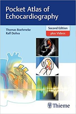
 Authors: Thomas Boehmeke, MD and Ralf Doliva, MD
Publisher: Thieme – 225 pages, with 444 illustrations
Book Review by: Nano Khilnani
Color Doppler echocardiography makes it easier for the physician, especially those who are new, to interpret what they see. A lot of important data is presented with this imaging modality. ”Learning how to use this fascinating tool however is complicated,” the author Dr. Thomas Boehmeke points out in his Preface to the first edition that came out in 2015. So this book was published as a guide to interpreting color Doppler imaging with ease..
This compact (7.5”x 5”) book that fits in a coat pocket, is an excellent visual guide for use in ‘on-the-go’ diagnostic cardiology. Almost the entire length of the book consists of two images per page with captions below those images. So it is actually a picture book that enables you to quickly glance at an echocardiogram and interpret the results.
This mainly visual book, a second edition published in 2018, consists of Doppler images and full-color drawings. Below we list the parts of this book to provide you as an overview, what you will find in it. They are not entire chapters. They are one to two pages in a sort of mini section beginning with a heading, with two to four images pertaining to that heading.
Examination
- Imaging and Patient Position
- Parasternal Long-Axis View
- Parasternal Short-Axis View
- Apical Windows
- Suprasternal Window
- Subcostal Window
M-Mode and Doppler Echocardiography
- M-Mode Echocardiography
- Doppler Echocardiography
Cardiac Abnormalities
- Valvular Heart Disease
- Coronary Heart Disease
- Cardiomyopathies
- Prosthetic Valves
- Carditis
- Septal Defects
- Hypertensive Heart Diseases
- Intracardiac Masses
Video Contents:
- 3D Echocardiography
- Normal Findings
- Abnormal Findings of the Left Ventricle
- Mitral Valve
- Aortic Valve
- Tumor in the Left Atrium
- Pulmonary Hypertension
- Pathological Changes in the Right Atrium and Atrial Septum
- Aortic Dissection
- Pericardial Effusion
- Prosthetic Heart Valves
- Trans-esophageal Echocardiography
- Stress Echocardiography
Watch 79 videos online by getting the access code on the inside front cover of this book and going to www.MediaCenter.Thieme.com.
The major features of this book:
- A practical overview of the typical patient examination including imaging and patient positioning
- All cardiac diseases are shown: cardiomyopathies, carditis, coronary heart disease, hypertensive heart diseases, intracardiac masses: depicted in B-mode, M-mode, Doppler and color Doppler, prosthetic valves, septal defects, and valvular heart disease
- Detailed descriptions of all the acoustic windows and imaging planes for every echocardiogram
- More than 400 illustrations, including sharp, clear echocardiograms, corresponding fill-color schematic diagrams, and 3-D images. For this new edition, all images showing cardiac diseases have been processed to make them clearer and in more accurate elation to the explanatory drawings
- New to the second edition: nearly 80 videos are now available in the Thieme Media Center showing normal views and many pathologies in video mode
This is a nifty little echocardiography guide that can turn out to be quite important when evaluating a patient under time pressure. It is a good, practical contribution to cardiology practice in today’s fast-paced world.
Authors:
Thomas Boehmeke, MD is a cardiology specialist in private practice in Gladbeck, Germany
Ralf Doliva, MD is senior physician at the Cardiology at Clinic at Marienhospital Gelsenkirchen in Gelsenkirchen, Germany.


