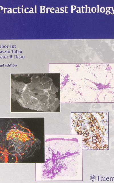
 Authors: Tibor Tot, MD; Lasazlo Tabar, MD; and Peter B. Dean, MD
Publisher: Thieme – 198 pages; 521 illustrations
Book Review by: Nano Khilnani
This book, with more than 500 images, takes an up-to-date, modern, multidisciplinary look at diagnosing, treating, and managing diseases of the breast.
The basic principles, radiologic appearance, and the pathology of breast disease are well laid out in this excellent book. Non-pathologists are not overwhelmed with excessive pathologic details.
This book provides clear information, plenty of case examples, and outstanding full-color and black-and-white images with detailed captions that can help you the breast disease specialist or trainee, the tools you need to do your work, or learn.
The images are not only what is found looking through microscope, but also photographs of actual diseased tissues, with the instruments used for examination and rulers that measure sizes. In addition, ultrasound images and MRIs are also used throughout this book.
The three authors of this volume of almost two hundred pages have produced something that provides a good overview of various diseases of the breast, with these subjects covered in its Contents:
- Normal Breast Tissue or Fibrocystic Change?
- General Morphology of Breast Lesions
- Hyperplastic Changes with and without Atypia
- Ductal Carcinoma in Situ
- The Most Common Types of Invasive Breast Carcinoma
- The Most Benign Breast Lesions and their Borderline and Malignant Counterparts
- Fine-Needle Aspiration or Core Biopsy: A Preoperative Diagnostic Algorithm
- The Postoperative Workup
- Assessment of the Most Important Prognostic Parameters
- Case Reports
This book is useful for practitioners – breast surgeons, oncologists, pathologists, and radiologists – as well as medical residents and students.
The material in each of the chapters is organized with sections such as:
- Diagnostic Criteria (laid out with bullet points)
- Differential Diagnosis (laid out with numbers)
- Conclusions
- Bibliography
In addition, for systematic absorption of material, the book has charts and tables with data.
Among the various benefits of using this book (second edition) are the following:
- High-quality examples of modern multimodality radiology (digital mammography, ultrasonography and magnetic resonance imaging or MRIs correlated with large format two-dimensional and three-dimensional histologic slides
- New findings on such clinically important topics as the lobar nature of breast carcinoma, multifocality, diffuse carcinomas and extent of disease, and concept of the sick lobe
- Introduction of the molecular classification of invasive breast cancer
- Widened discussion of key prognostic and predictive factors in breast carcinoma, such as hormone receptors and HER2 status
- Updates on preoperative diagnosis, including intact biopsy and radiologic assessment of the extent and distribution of lesions.
Authors:
TiborTot, MD is Associate Professor of the Department of Pathology and Clinical Cytology at Falun Central Hospital in Falun, Switzerland.
Laszlo Tabar, MD is Professor Emeritus in the Department of Mammography at Falun Central Hospital in Falun, Switzerland.
Peter B. Dean, MD is Professor Emeritus in the Department of Diagnostic Radiology at University Central Hospital in Turku, Finland; and Former Director of Breast Imaging there.


