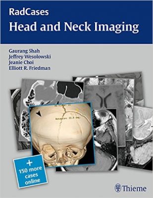
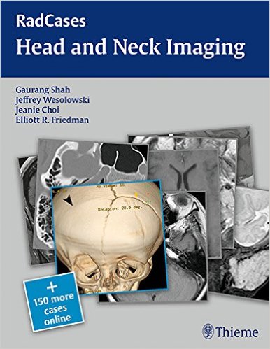
 Editors: Gaurang Shah, MD; Jeffrey Wesolowski, MD; Jeanie Choi, MD; and Elliott R. Friedman, MD.
Series Editors: Jonathan Lorenz, MD and Hector Ferral, MD.
Publisher: Thieme – 213 pages, with 100 carefully-selected cases. Plus 150+ cases online.
Book Review by: Nano Khilnani
While this book has been written for you the radiology trainee and resident to review what you learned in medical school, as well as take a look at the cases presented in it to prepare of the American Board of Radiology examination, it’s also a great resource you can go back to again and again to do closer analyses, and get deeper understanding of abnormalities you find in the cases you see.
This volume is also a valuable asset to the established radiologist, otolaryngologist, and head and neck surgeon. The editors write that “practitioners in this field are some of the finest diagnosticians in medicine. They have to be. And the best way to prepare for a career as a head and neck specialist is to study all aspects associated with the field so that you can develop your skills during training and on the job.”
Radiographs are used not only in head and neck cases, but in most medical specialties to check for diseases and disorders. “Radiology stands at the forefront of medical analysis…,” write Drs. Shah, Wesolowski, Choi, and Friedman in their Preface.
In this book you will find a wide variety of patient presentations. And there are more cases available online by going to www.MediaCenter.Thieme.com. Simply scratch off the access code on the inside front cover of this book and enter it when you register your copy.
The series editors, Drs. Lorenz and Ferral, point out that “despite ongoing efforts to improve Web-based interactive databases, residents still find themselves searching for material they can review while preparing for the radiology board examinations and remain frustrated by the fact that only a few printed guides are available, which are limited in both format and image quality.”
The main purpose for creating this series of books called RadCases was to combine the frequent and wide use printed books with the “adaptability, exceptional quality, and the interactive features of an electronic, case-based format.” This printed book enables wider use by presenting a set of exceptional core (‘must-know’) cases that the resident can get very familiar with and eventually master.
This book is easier to use than others because it is provided with annotations and descriptions for all images. Also, the search engine available on the website indicated above, enables you to create individualized, daily study lists that are not limited by factors such as radiology subsection. So you can for example, study tuberculosis in every subspecialty and in any system of the body.
As another example, you can study only thoracic cases, including those with links to say, cardiology and/or nuclear medicine, and/or pediatrics. You want to study only musculoskeletal cases? You can do so if you wish – the choice is yours.
This book covers: breast imaging, cardiac imaging, emergency radiology, gastrointestinal imaging, genitourinary imaging, head and neck imaging, interventional radiology, musculoskeletal radiology, neuro-imaging, nuclear medicine, pediatric imaging, thoracic imaging, and ultrasound imaging.
The 100 cases featured in this book have been carefully selected by the editors and contain clear, high-quality radiographs. They are also well organized so that you can get maximum ease of use for assessment by yourself.
Each case starts with the clinical presentations on the right-side page. Simply turn the page for image findings, differential diagnoses, the definitive diagnosis, essential facts, and more.
Editors:
Gaurang Shah, MD is Associate Professor in the Department of Radiology at the University of Michigan in Ann Arbor, Michigan.
Jeffrey Wesolowski, MD is Assistant Professor and Associate Neuroradiology Division Director in the Department of Radiology at the University of Michigan in Ann Arbor, Michigan.
Jeanie Choi, MD is Assistant Professor in the Department of Diagnostic and Interventional Imaging at McGovern Medical School at the University of Texas Health Science Center in Houston, Texas.
Elliott R. Friedman, MD is Assistant Professor in the Department of Diagnostic and Interventional Imaging at McGovern Medical School at the University of Texas Health Science Center in Houston, Texas.


