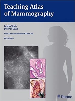
 Authors: Laszlo Tabar, MD; and Peter W. Dean, MD;
Contributor: Tibor Tot, MD
Publisher: Thieme – 300 pages, with 669 illustrations
Book Review by: Nano Khilnani
Teaching you – the breast specialist and / or radiologist – how to analyze mammograms and arrive at the correct diagnosis is the main objective of the authors of this book.
Many of the images used in this text are taken from actual patients’ records accumulated for a period of up to 25 years. They represent a wide range, variety, and history of abnormalities found upon examining the breasts of women.
The authors write in their Introduction that mammography seeks principally to prevent the growth of breast cancers in women, and one way to do this is, is to enable you the clinician to detect subtle hints of malignancy when tiny and / or obscure structures are found.
Textbooks on mammography typically show different types and stages of breast lesions after they were already found. This book is different. It is designed to assist you in learning how to detect abnormal or out-of the-ordinary patterns such as architectural distortions found in mammograms, closely analyze the images, and reach the right diagnosis.
Many series of mammograms are shown to you the reader. Many have tiny traces of something, like a blur, or an unusual pattern. Through frequent observations and constant practice in pinpointing abnormalities, you can greatly increase your chances of success in detection with the aid of this book.
The authors point out that the “prerequisites for the perception and evaluation of mammographic findings” are:
- Optimum technique
- Knowledge of anatomy
- Understanding of the pathological processes leading to the mammographic appearances
Replete with numerous full-color micrographs, black-and-white radiographs, some actual photos of breast tissue, and diagrams, this book has a wealth of information on mammogram analysis as well as on various imaging modalities. It contains eight chapters, namely:
- Anatomy of the Breast
- Method for Systematic Viewing of Mammograms
- Approach to Viewing Mammographic Images and Interpreting the Findings
- Circular / Oval Lesions
- Stellate / Spiculated Lesions and Architectural Distortion
- Calcifications on the Mammogram
- Thickened Skin Syndrome of the Breast
- Overall Strategy
One of the most important of these is chapter 2, Method for Systematic Viewing of Mammograms. It provides a good grounding on how to look at mammograms using a systematic method every time.
The authors start out this chapter by stating: “A detailed comparison of the left and right breasts enhances the detection of structural asymmetries. Perception of subtle alterations can be accentuated by using masking techniques to enable sequential viewing of restricted areas of the mammograms.”
“Masking is equally necessary for viewing film or digital formats. A hand-held viewer is an excellent tool for viewing films viewer (Fig. X-II). Digital software can perform this function when viewing soft-copy images on digital monitors.”
The authors show images of two types of masking – horizontal and oblique – and inform us that the goal of perception is to:
- Find asymmetrical densities
- Examine architectural distortion
- Detect parenchymal contour
- See calcification on the mammogram
Using masking techniques, the authors show mammograms in the pages of this chapter that reveal some abnormalities found in the breasts, such as a radiating structure, a small stellate lesion, and a carcinoma that may otherwise have not been found using other means of detection.
In chapter 3, Approach to Viewing Mammographic Images and Interpreting the Findings, the authors inform us that when interpreting mammograms, these three important steps should be taken:
- Determine whether the image is of diagnostic quality
- Perform a systematic, step-by-step survey of the mammogram to evaluate details of the breast structure while searching for a lesion
- Carefully analyze each detected lesion
- Then, place each lesion into one of these five classifications:
- Circulo/oval lesions that may be solitary or multiple
- Stellate/speculated lesions and architectural distortion
- Calcifications that may or not be associated with any tumor
- Thickened skin syndrome
- Any combination of two or more of the above types
After such classification, each detected lesion should undergo detailed analysis, as described in chapters 4 through 7.
This is an excellent textbook on mammography, with a focus on teaching tried and tested, systematic and successful methods.
Editors:
Laszlo Tabar, MD is Professor of Radiology at the University of Uppsala School of Medicine, and Medical Director of the Department of Mammography at Falun Central Hospital in Falun, Sweden.
Peter W. Dean, MD is Professor in the Department of Diagnostic Radiology at the University of Turku, and Director of Breast Imaging at Turku University Hospital in Turku, Finland.
Conributor:
Tibor Tot, MD, PhD is Associate Professor of Pathology, and Chairman of the Department of Pathology and Clinical Cytology at Central Hospital in Falun, Sweden.


