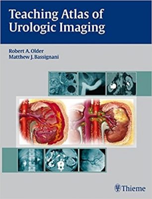
 Editors: Robert A. Older, MD; and Matthew J. Bassignani, MD
Publisher: Thieme – 350 pages, with nearly 400 high-quality illustrations, 47 in full-color
Book Review by: Nano Khilnani
The format of this book is vastly different from others. Instead of chapters, the contents are presented in the form of cases. A brief clinical history is given in each case, along with an image or series of images. This is similar to learning in hospitals where residents make the rounds visiting patients, and are asked questions by attending physicians.
Here are some steps the editors present in the learning process:
- The case can be initially viewed as an unknown
- After viewing images, the student can view the pertinent radiographic findings
- The correct diagnosis is then provided
- Then, a brief differential diagnosis of other entities that might be considered given the imaging findings, follows
- Clinical and pathologic data and treatment options are presented where appropriate
- The Imaging Findings section that follows is meant to:
- Discuss reasons why other choices in the differential diagnosis list are not correct
- Discuss the role of imaging modalities other than the imaging study used for the initial case discussion. Sometimes this includes many modalities, but for other cases only limited modalities are appropriate.
- Diagnostic ‘pearls’ are included as appropriate
- A Suggested Readings list is provided at the end of each case presentation.
A total of 93 cases are provided and discussed in this book of 350 pages, which are organized around 12 Parts we name below to give you a broad overview:
- Part I – Congenital Abnormalities
- Part II – Contrast Media
- Part III – Stone Disease, Obstruction, and Metabolic Abnormalities
- Part IV – Urinary Tract Infection
- Part V – Renal Cystic Disease
- Part VI – Solid Renal Neoplasms
- Part VII – Transitional Cell Carcinomas and Filling Defects
- Part VIII – Urinary Tract Trauma / Upper and Lower Urinary Tract
- Part IX – Adrenal Abnormalities
- Part X – Vascular Abnormalities
- Part XI – Testicular and Scrotal Abnormalities
- Part XII – Miscellaneous
Let’s take a look at the outline of the presentation of Case 21 in Part IV, Urinary Tract Infection:
Clinical Presentation
- A 27-year-old male presents with left flank pain and low-grade fever.
- Figure 21.1 shows two computed tomography (CT) scans at two levels through the left kidney
Radiologic Findings
- Focal hypodense area (Fig.21.1A
- Striated nephrogram (arrowheads, Fig.21.1B)
- Slight nephrographic delay in function for the left kidney (Fig.21.1A,B)
Diagnosis
- Acute pyelonephritis (AP)
Differential Diagnosis
- Renal tumor
- Ureteral obstruction
- Contusion
Discussion
Background
Most AP results from an ascending urinary tract infection (UTI). The most common organism is Escherichia coli (8o percent) in adults. A minority of AP cases are caused by hematogenous spread from distant sources (e.g. endocarditis, tooth abscess, intravenous drug abuse with septic thrombophlebitis). Most common such organisms are Staphylococcus and Streptococcus.
Clinical Findings
A discussion of what acute pyelonephritis is, its symptoms such as flank pain and fever; results of blood analysis such as elevated white blood cell count; evidence of upper tract infection; recommendation for further examination through renal imaging only if diagnosis is in doubt or if patient is unresponsive to appropriate antibiotic coverage beyond 72 hours.
Pathology
In uncomplicated AP, imaging findings (e.g. poor enhancement, striated nephrogram) are attributed to interstitial edema and / or collecting tubules clogged with inflammatory cells, debris, or both.
Imaging Findings
- Computed Tomography
- Radiography
- Ultrasound
- Nuclear Medicine
Treatment
Uncomplicated AP responds well to antimicrobial therapy.
Prognosis
In the absence of recurrent disease with scarring, the prognosis is excellent.
Pearls
All UTIs in male children and females under five years old require imaging to evaluate for vesicoureteral reflux and other predisposing conditions
Suggested Readings
Three sources of information are listed.
The case-based format of this book makes it a valuable reference source for radiology, urology, and nephrology residents, especially those preparing for board examinations. This is also useful to established practitioners who want to look at the range of findings from imaging.
In it the editors suggest the best imaging methods for different types of diseases, disorders, and conditions. All in all, this is an excellent volume that provides ample information: lots of bulleted lists, great illustrations, and much more.
Editors:
Robert A. Older, MD is Professor Emeritus in the Department of Radiology at the University of Virginia Health Sciences Center in Charlottesville, Virginia; and Director of Imaging at Virginia Urology Center in Richmond, Virginia.
Matthew J. Bassignani, MD is Associate Professor in the Department of Radiology, Section Chief of Genitourinary Imaging, Medical Director for Radiology Information Systems, and Medical Director of the University of Virginia Imaging Center at the University of Virginia Health Sciences Center in Charlottesville, Virginia.


