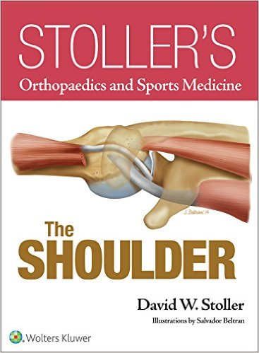
 (Part of Stoller’s Orthopedic and Sports Medicine series that includes the elbow, the foot and ankle, the hip, the knee, and the wrist and hand)
Editor: David W. Stoller, MD; Illustrator: Salvador Beltran
Publisher: Wolters Kluwer | Lippincott, Williams & Wilkins – 862 pages
Book Review by: Hiro Motiram
This big, bright, well-illustrated book on the shoulder should become an important part of the library of practicing and aspiring orthopedists including orthopedic surgeons such as you for example, if it is not already occupying a central space in your collection.
This massive volume of work represents an outstanding achievement of Dr. David W. Stoller, a notable, distinguished orthopedic surgeon who as editor, compiled, organized, and presented the materials of four ‘shoulder experts’ as well as his own, to present to you for your practice. For a book of this size, one would expect a lot of chapters and perhaps be foreboding to read, but the number of chapters – just eight – is refreshingly welcome, especially to medical residents and students who are used to huge and lengthy tomes.
To give you an overview of what you will find in this highly useful book, we present to you below the titles of the nine chapters of this book of 862 pages:
- Practical Guide to Shoulder MR Imaging
- Shoulder Anatomy
- Stoller’s Shoulder Checklist and Protocols
- Glenohumeral Joint and Capsular Gross Anatomy
- Shoulder Impingement Syndrome
- Rotator Cuff Tears, Microinstability, Rotator Cuff Interval/Biceps Pulley, and the Throwing Shoulder
- Glenohumeral Joint Instability
- Inferior Glenohumeral Ligament Capsular Variations, High Attachment Anterior Band, and SLAP Lesions
- Shoulder- related Pathology Including Paralabral Cysts and Biceps Tendon
Ample use of large-print text, various types of images including arthrograms, arthroscopic views, full-color line drawings and photos, and other visual items, help you to easily view and learn the nature of the ailment, injury, lesion, disease, disorder, tear or other condition. This book very systematically provides steps in text form on how to best relieve a condition, repair a tear, or take other surgical actions, along with accompanying images.
Among the notable features of this book are the following:
- 3T and high-resolution magnetic resonance (MR) images that demonstrate critical structures in functional shoulder anatomy and pathology
- Comprehensive collections of color illustrations and arthroscopic cases of orthopedic pathoanatomy
- Detailed figure legends rich in content that provide descriptive information and introduce novel concepts
- Evolved checklist approach as the keystone for accurate and reproducible image interpretation
- Key concepts section introductions that emphasize and reinforce critical information
- Updated concepts on the shoulder including:
- Cadaver dissections highlighting anterior band and capsular variations
- Partial articular supraspinatus tendon avulsion (PASTA) lesions in the context of rotator cable anatomy
- Proper cuff tensioning in rotator cuff repair
- Rotator cable
- Superior glenohumeral ligament complex (SGLC), inferior glenohumeral ligament labral complex (IGLLC) and biceps labral complex (BLC).
This well-organized book is an excellent source of information on all matters concerning the human shoulder, and is a good guide to relevant anatomy, pathology, and surgery.
Editor:
David W. Stoller, MD, FACR is National Director of Orthopedic and Musculoskeletal Imaging at RadNet; Medical Director of Orthopedic and Musculoskeletal Imaging at Beverly Radiology, Northern California, in San Francisco, California; and Affiliate Member of American Shoulder and Elbow Surgeons.
Contributors:
Lesley J. Anderson, MD is Assistant Clinical Professor of Orthopedic Surgery at University of California, San Francisco; and Orthopedic Surgeon at California Pacific Medical Center in San Francisco, California.
Paul B. Roache, MD is an orthopedic surgeon at California Pacific Medical Center in San Francisco, California.
David W. Stoller, MD, FACR (see above).
Eugene M. Wolf, MD is with the Department of Orthopedic Surgery at St. Mary’s Medical Center in San Francisco, California.
Scientific-Research Contributors:
Sophia D. Heber, MD is with Johannes Gutenberg, University of Maine.
David Hitt, MSc, RT (R), MR is MR Clinical Adoption Specialist at U.S. MR Marketing Department at Philips Healthcare.
Adriana Kanwicher, BS is with Medical Modality, UNISA; Advanced Applications Specialist, Global MR; and GE Healthcare Technologies, Magnetic Resonance.
Robert D. Peters, PhD is Manager of Global MR MSK; Applications & Physics, Core Technologies; and GE Healthcare Technologies, Magnetic Re


